Click image to see more details
Product Info Summary
| SKU: | A04441-3 |
|---|---|
| Size: | 100 μg/vial |
| Reactive Species: | Human, Mouse |
| Host: | Rabbit |
| Application: | ELISA, Flow Cytometry, WB |
Customers Who Bought This Also Bought


Product info
Product Name
Anti-GIT2 Antibody Picoband®
SKU/Catalog Number
A04441-3
Size
100 μg/vial
Form
Lyophilized
Description
Boster Bio Anti-GIT2 Antibody Picoband® catalog # A04441-3. Tested in ELISA, Flow Cytometry, WB applications. This antibody reacts with Human, Mouse. The brand Picoband indicates this is a premium antibody that guarantees superior quality, high affinity, and strong signals with minimal background in Western blot applications. Only our best-performing antibodies are designated as Picoband, ensuring unmatched performance.
Storage & Handling
At -20°C for one year from date of receipt. After reconstitution, at 4°C for one month. It can also be aliquotted and stored frozen at -20°C for six months. Avoid repeated freezing and thawing.
Cite This Product
Anti-GIT2 Antibody Picoband® (Boster Biological Technology, Pleasanton CA, USA, Catalog # A04441-3)
Host
Rabbit
Contents
Each vial contains 4 mg Trehalose, 0.9 mg NaCl, 0.2 mg Na2HPO4.
Clonality
Polyclonal
Isotype
Rabbit IgG
Immunogen
E.coli-derived human GIT2 recombinant protein (Position: D400-S556).
*Blocking peptide can be purchased. Costs vary based on immunogen length. Contact us for pricing.
Cross-reactivity
No cross-reactivity with other proteins.
Reactive Species
A04441-3 is reactive to GIT2 in Human, Mouse
Reconstitution
Adding 0.2 ml of distilled water will yield a concentration of 500 μg/ml.
Observed Molecular Weight
85 kDa
Calculated molecular weight
84.543kDa
Background of GIT2
ARF GTPase-activating protein GIT2 is an enzyme that in humans is encoded by the GIT2 gene. This gene encodes a member of the GIT protein family, which interact with G protein-coupled receptor kinases and possess ADP-ribosylation factor (ARF) GTPase-activating protein (GAP) activity. GIT proteins traffic between cytoplasmic complexes, focal adhesions, and the cell periphery, and interact with Pak interacting exchange factor beta (PIX) to form large oligomeric complexes that transiently recruit other proteins. GIT proteins regulate cytoskeletal dynamics and participate in receptor internalization and membrane trafficking. This gene has been shown to repress lamellipodial extension and focal adhesion turnover, and is thought to regulate cell motility. This gene undergoes extensive alternative splicing to generate multiple isoforms, but the full-length nature of some of these variants has not been determined. The various isoforms have functional differences, with respect to ARF GAP activity and to G protein-coupled receptor kinase 2 binding.
Antibody Validation
Boster validates all antibodies on WB, IHC, ICC, Immunofluorescence, and ELISA with known positive control and negative samples to ensure specificity and high affinity, including thorough antibody incubations.
Application & Images
Applications
A04441-3 is guaranteed for ELISA, Flow Cytometry, WB Boster Guarantee
Assay Dilutions Recommendation
The recommendations below provide a starting point for assay optimization. The actual working concentration varies and should be decided by the user.
Western blot, 0.1-0.25 μg/ml, Human, Mouse
Flow Cytometry (Fixed), 1-3 μg/1x106 cells, Human
ELISA, 0.1-0.5 μg/ml, Human
Positive Control
WB: human Hela whole cell, human Jurkat whole cell, human HepG2 whole cell, mouse lung tissue
FCM: PC-3 cell
Validation Images & Assay Conditions
Click image to see more details
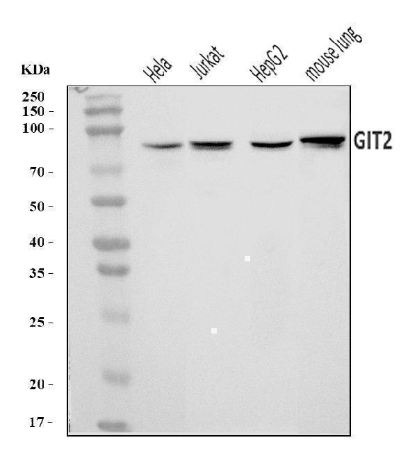
Figure 1. Western blot analysis of GIT2 using anti-GIT2 antibody (A04441-3).
Electrophoresis was performed on a 5-20% SDS-PAGE gel at 70V (Stacking gel) / 90V (Resolving gel) for 2-3 hours. The sample well of each lane was loaded with 30 ug of sample under reducing conditions.
Lane 1: human Hela whole cell lysates,
Lane 2: human Jurkat whole cell lysates,
Lane 3: human HepG2 whole cell lysates,
Lane 4: mouse lung tissue lysates.
After electrophoresis, proteins were transferred to a nitrocellulose membrane at 150 mA for 50-90 minutes. Blocked the membrane with 5% non-fat milk/TBS for 1.5 hour at RT. The membrane was incubated with rabbit anti-GIT2 antigen affinity purified polyclonal antibody (Catalog # A04441-3) at 0.25 μg/mL overnight at 4°C, then washed with TBS-0.1%Tween 3 times with 5 minutes each and probed with a goat anti-rabbit IgG-HRP secondary antibody at a dilution of 1:5000 for 1.5 hour at RT. The signal is developed using an Enhanced Chemiluminescent detection (ECL) kit (Catalog # EK1002) with Tanon 5200 system. A specific band was detected for GIT2 at approximately 85 kDa. The expected band size for GIT2 is at 85 kDa.

Click image to see more details
Figure 2. Flow Cytometry analysis of PC-3 cells using anti-GIT2 antibody (A04441-3).
Overlay histogram showing PC-3 cells stained with A04441-3 (Blue line). To facilitate intracellular staining, cells were fixed with 4% paraformaldehyde and permeabilized with permeabilization buffer. The cells were blocked with 10% normal goat serum. And then incubated with rabbit anti-GIT2 Antibody (A04441-3, 1 μg/1x106 cells) for 30 min at 20°C. DyLight®488 conjugated goat anti-rabbit IgG (BA1127, 5-10 μg/1x106 cells) was used as secondary antibody for 30 minutes at 20°C. Isotype control antibody (Green line) was rabbit IgG (1 μg/1x106) used under the same conditions. Unlabelled sample without incubation with primary antibody and secondary antibody (Red line) was used as a blank control.
Protein Target Info & Infographic
Gene/Protein Information For GIT2 (Source: Uniprot.org, NCBI)
Gene Name
GIT2
Full Name
ARF GTPase-activating protein GIT2
Weight
84.543kDa
Alternative Names
Interleukin-25; IL-25; Interleukin-17E; IL-17E; IL25; IL17E; UNQ3120/PRO10272 GIT2 CAT-2, CAT2, PKL GIT ArfGAP 2 ARF GTPase-activating protein GIT2|ARF GAP GIT2|G protein-coupled receptor kinase interacting ArfGAP 2|G protein-coupled receptor kinase-interactor 2|GRK-interacting protein 2|Paxillin kinase linker|cool-associated, tyrosine phosphorylated protein 2|cool-interacting tyrosine-phosphorylated protein 2
*If product is indicated to react with multiple species, protein info is based on the gene entry specified above in "Species".For more info on GIT2, check out the GIT2 Infographic

We have 30,000+ of these available, one for each gene! Check them out.
In this infographic, you will see the following information for GIT2: database IDs, superfamily, protein function, synonyms, molecular weight, chromosomal locations, tissues of expression, subcellular locations, post-translational modifications, and related diseases, research areas & pathways. If you want to see more information included, or would like to contribute to it and be acknowledged, please contact [email protected].
Specific Publications For Anti-GIT2 Antibody Picoband® (A04441-3)
Hello CJ!
No publications found for A04441-3
*Do you have publications using this product? Share with us and receive a reward. Ask us for more details.
Recommended Resources
Here are featured tools and databases that you might find useful.
- Boster's Pathways Library
- Protein Databases
- Bioscience Research Protocol Resources
- Data Processing & Analysis Software
- Photo Editing Software
- Scientific Literature Resources
- Research Paper Management Tools
- Molecular Biology Software
- Primer Design Tools
- Bioinformatics Tools
- Phylogenetic Tree Analysis
Customer Reviews
Have you used Anti-GIT2 Antibody Picoband®?
Submit a review and receive an Amazon gift card.
- $30 for a review with an image
0 Reviews For Anti-GIT2 Antibody Picoband®
Customer Q&As
Have a question?
Find answers in Q&As, reviews.
Can't find your answer?
Submit your question
