
Click image to see more details
Product Info Summary
| SKU: | M00399 |
|---|---|
| Size: | 100ug/vial |
| Reactive Species: | Human |
| Host: | Mouse |
| Application: | Flow Cytometry, IF, WB |
Customers Who Bought This Also Bought

Product info
Product Name
Anti-FLI1 (Ewing s Sarcoma & Endothelial Marker) Monoclonal Antibody
SKU/Catalog Number
M00399
Size
100ug/vial
Form
Liquid
Description
Boster Bio Anti-FLI1 (Ewing s Sarcoma & Endothelial Marker) Monoclonal Antibody (Catalog # M00399). Tested in Flow Cytometry, IF, WB applications. This antibody reacts with Human.
Storage & Handling
Antibody with azide - store at 2 to 8°C. Antibody without azide - store at -20 to -80°C. Antibody is stable for 24 months. Non-hazardous. No MSDS required.
Cite This Product
Anti-FLI1 (Ewing s Sarcoma & Endothelial Marker) Monoclonal Antibody (Boster Biological Technology, Pleasanton CA, USA, Catalog # M00399)
Host
Mouse
Contents
Prepared in 10mM PBS with 0.05% BSA & 0.05% azide. Also available WITHOUT BSA at 1.0mg/ml.
Clonality
Monoclonal
Clone Number
Clone: FLI1/1312
Isotype
IgG1, kappa
Immunogen
Recombinant fragment (aa 107-261) of human FLI1 protein with hexa-histidine tag
*Blocking peptide can be purchased. Costs vary based on immunogen length. Contact us for pricing.
Reactive Species
M00399 is reactive to FLI1 in Human
Reconstitution
Reconstitute with 1ml water (15 minutes at room temperature).
Calculated molecular weight
50982 MW
Background of FLI1
Recognizes a protein of 51kDa, which is identified as FLI1. This protein, a member of the ETS family of DNA binding transcription factors, is involved in cellular proliferation and tumorigenesis. Ets-1 is the prototype member of a family of genes identified on the basis of homology to the v-Ets oncogene isolated from the E26 erythroblastosis virus.Members of the Ets gene family share a highly conserved carboxy-terminal domain containing a sequence related to the SV40 large T antigen nuclear localization signal sequence. Approximately 90% of Ewing's Sarcoma (EWS) / Primitive Neuroectodermal Tumors (PNET) have a specific translocation, t(11;22)(q24;q12), which results in fusion of EWS to Fli-1, and production of an EWS-Fli-1 fusion protein. Among normal tissues only endothelial cells and small lymphocytes express Fli-1. This protein is expressed in majority of vascular tumors including angiosarcomas, hemangioendotheliomas, hemangiomas, and Kaposi's Sarcomas. High sensitivity and specificity of Fli-1 equals to or exceeds that of the established vascular markers like CD31, CD34, and Factor VIII.
Antibody Validation
Boster validates all antibodies on WB, IHC, ICC, Immunofluorescence, and ELISA with known positive control and negative samples to ensure specificity and high affinity, including thorough antibody incubations.
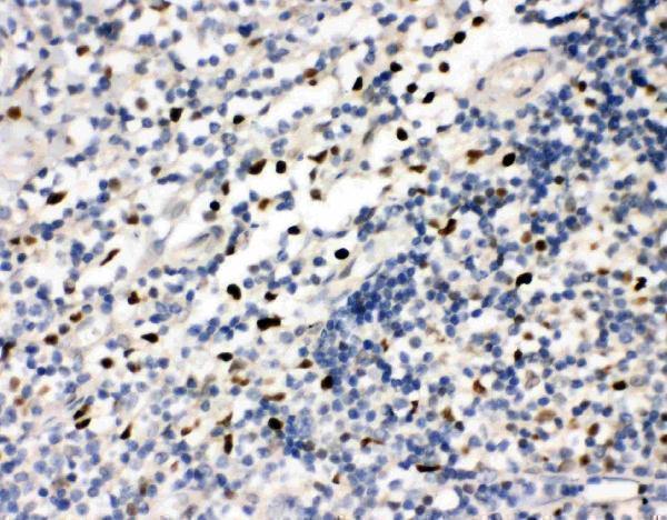
Application & Images
Applications
M00399 is guaranteed for Flow Cytometry, IF, WB Boster Guarantee
Assay Dilutions Recommendation
The recommendations below provide a starting point for assay optimization. The actual working concentration varies and should be decided by the user.
Flow Cytometry (1-2ug/million cells)
Immunofluorescence (1-2ug/ml)
Western Blot (1-2ug/ml for 60 minutes at RT)
Optimal dilution for a specific application should be determined.
Validation Images & Assay Conditions
Click image to see more details
Western Blot of THP1 and Raji cell lysate using Anti-FLI1 Monoclonal Antibody (FLI1/1312)
Protein Target Info & Infographic
Gene/Protein Information For FLI1 (Source: Uniprot.org, NCBI)
Gene Name
FLI1
Full Name
Friend leukemia integration 1 transcription factor
Weight
50982 MW
Superfamily
ETS family
Alternative Names
Friend leukemia integration 1 transcription factor;Proto-oncogene Fli-1;Transcription factor ERGB;FLI1; FLI1 BDPLT21, EWSR2, SIC-1 Fli-1 proto-oncogene, ETS transcription factor Friend leukemia integration 1 transcription factor|Ewing sarcoma breakpoint region 2|Friend leukemia virus integration 1|transcription factor ERGB
*If product is indicated to react with multiple species, protein info is based on the gene entry specified above in "Species".For more info on FLI1, check out the FLI1 Infographic

We have 30,000+ of these available, one for each gene! Check them out.
In this infographic, you will see the following information for FLI1: database IDs, superfamily, protein function, synonyms, molecular weight, chromosomal locations, tissues of expression, subcellular locations, post-translational modifications, and related diseases, research areas & pathways. If you want to see more information included, or would like to contribute to it and be acknowledged, please contact [email protected].
Specific Publications For Anti-FLI1 (Ewing s Sarcoma & Endothelial Marker) Monoclonal Antibody (M00399)
Hello CJ!
No publications found for M00399
*Do you have publications using this product? Share with us and receive a reward. Ask us for more details.
Recommended Resources
Here are featured tools and databases that you might find useful.
- Boster's Pathways Library
- Protein Databases
- Bioscience Research Protocol Resources
- Data Processing & Analysis Software
- Photo Editing Software
- Scientific Literature Resources
- Research Paper Management Tools
- Molecular Biology Software
- Primer Design Tools
- Bioinformatics Tools
- Phylogenetic Tree Analysis
Customer Reviews
Have you used Anti-FLI1 (Ewing s Sarcoma & Endothelial Marker) Monoclonal Antibody?
Submit a review and receive an Amazon gift card.
- $30 for a review with an image
0 Reviews For Anti-FLI1 (Ewing s Sarcoma & Endothelial Marker) Monoclonal Antibody
Customer Q&As
Have a question?
Find answers in Q&As, reviews.
Can't find your answer?
Submit your question
4 Customer Q&As for Anti-FLI1 (Ewing s Sarcoma & Endothelial Marker) Monoclonal Antibody
Question
We were content with the WB result of your anti-FLI1 (Ewing s Sarcoma & Endothelial Marker) Monoclonal antibody. However we have observed positive staining in placenta nucleus. using this antibody. Is that expected? Could you tell me where is FLI1 supposed to be expressed?
T. Huang
Verified customer
Asked: 2019-09-26
Answer
Based on literature, placenta does express FLI1. Generally FLI1 expresses in nucleus. Regarding which tissues have FLI1 expression, here are a few articles citing expression in various tissues:
Amygdala, Placenta, and Thymus, Pubmed ID: 14702039
Blood, Pubmed ID: 8439553
Bone marrow, Pubmed ID: 1522903
Leukemic T-cell, Pubmed ID: 19690332
Lymph, Pubmed ID: 15489334
Placenta, Pubmed ID: 7542907
Boster Scientific Support
Answered: 2019-09-26
Question
you antibody using your anti-FLI1 (Ewing s Sarcoma & Endothelial Marker) Monoclonal antibody for animal organ morphogenesis studies. Has this antibody been tested with western blotting on raji cell lysate? We would like to see some validation images before ordering.
Verified Customer
Verified customer
Asked: 2019-06-19
Answer
We appreciate your inquiry. This M00399 anti-FLI1 (Ewing s Sarcoma & Endothelial Marker) Monoclonal antibody is tested on raji cell lysate. It is guaranteed to work for WB in human. Our Boster guarantee will cover your intended experiment even if the sample type has not been be directly tested.
Boster Scientific Support
Answered: 2019-06-19
Question
We are currently using anti-FLI1 (Ewing s Sarcoma & Endothelial Marker) Monoclonal antibody M00399 for human tissue, and we are satisfied with the WB results. The species of reactivity given in the datasheet says human. Is it likely that the antibody can work on primate tissues as well?
K. Anderson
Verified customer
Asked: 2017-04-04
Answer
The anti-FLI1 (Ewing s Sarcoma & Endothelial Marker) Monoclonal antibody (M00399) has not been validated for cross reactivity specifically with primate tissues, though there is a good chance of cross reactivity. We have an innovator award program that if you test this antibody and show it works in primate you can get your next antibody for free. Please contact me if I can help you with anything.
Boster Scientific Support
Answered: 2017-04-04
Question
We have seen staining in human amygdala. Are there any suggestions? Is anti-FLI1 (Ewing s Sarcoma & Endothelial Marker) Monoclonal antibody supposed to stain amygdala positively?
B. Kulkarni
Verified customer
Asked: 2016-11-04
Answer
From literature amygdala does express FLI1. From Uniprot.org, FLI1 is expressed in leukocyte, bone marrow, blood, amygdala, placenta thymus, lymph, placenta, leukemic t-cell, among other tissues. Regarding which tissues have FLI1 expression, here are a few articles citing expression in various tissues:
Amygdala, Placenta, and Thymus, Pubmed ID: 14702039
Blood, Pubmed ID: 8439553
Bone marrow, Pubmed ID: 1522903
Leukemic T-cell, Pubmed ID: 19690332
Lymph, Pubmed ID: 15489334
Placenta, Pubmed ID: 7542907
Boster Scientific Support
Answered: 2016-11-04
