Click image to see more details
-

-
-
-
-
+4
Product Info Summary
| SKU: | M01528-2 |
|---|---|
| Size: | 100 μg/vial |
| Reactive Species: | Human, Mouse, Rat |
| Host: | Mouse |
| Application: | IHC, WB |
Customers Who Bought This Also Bought


Product info
Product Name
Anti-FABP4 Antibody Picoband™ (monoclonal, 10E12)
View all FABP4/A-FABP Antibodies
SKU/Catalog Number
M01528-2
Size
100 μg/vial
Form
Lyophilized
Description
Boster Bio Anti-FABP4 Antibody Picoband™ (monoclonal, 10E12) catalog # M01528-2. Tested in IHC, WB applications. This antibody reacts with Human, Mouse, Rat.
Storage & Handling
At -20°C for one year from date of receipt. After reconstitution, at 4°C for one month. It can also be aliquotted and stored frozen at -20°C for six months. Avoid repeated freezing and thawing.
Cite This Product
Anti-FABP4 Antibody Picoband™ (monoclonal, 10E12) (Boster Biological Technology, Pleasanton CA, USA, Catalog # M01528-2)
Host
Mouse
Contents
Each vial contains 4 mg Trehalose, 0.9 mg NaCl and 0.2 mg Na2HPO4.
Clonality
Monoclonal
Clone Number
10E12
Isotype
Mouse IgG1
Immunogen
A synthetic peptide corresponding to a sequence at the N-terminus of human FABP4, identical to the related mouse and rat sequences.
*Blocking peptide can be purchased. Costs vary based on immunogen length. Contact us for pricing.
Cross-reactivity
No cross-reactivity with other proteins.
Reactive Species
M01528-2 is reactive to FABP4 in Human, Mouse, Rat
Applications
M01528-2 is guaranteed for IHC, WB Boster Guarantee
Observed Molecular Weight
15 kDa
Calculated molecular weight
Background of FABP4/A-FABP
Fatty acid binding proteins (FABPs) are small cytoplasmic proteins that are expressed in a highly tissue-specific manner and bind to fatty acids such as oleic and retinoic acid. Adipocyte fatty-acid-binding protein, aP2 (FABP4) is expressed in adipocytes and macrophages, and integrates inflammatory and metabolic responses. Studies in aP2-deficient mice have shown that this lipid chaperone has a significant role in several aspects of metabolic syndrome, including type 2 diabetes and atherosclerosis. It regulates allergic airway inflammation and may provide a link between fatty acid metabolism and asthma.
Antibody Validation
Boster validates all antibodies on WB, IHC, ICC, Immunofluorescence, and ELISA with known positive control and negative samples to ensure specificity and high affinity, including thorough antibody incubations.
Assay dilution & Images
Reconstitution
Adding 0.2 ml of distilled water will yield a concentration of 500 μg/ml.
Assay Dilutions Recommendation
The recommendations below provide a starting point for assay optimization. The actual working concentration varies and should be decided by the user.
Western blot, 0.25-0.5 μg/ml, Human, Mouse, Rat
Immunohistochemistry(Paraffin-embedded Section), 2-5 μg/ml, Human, Mouse, Rat
Validation Images & Assay Conditions
Click image to see more details
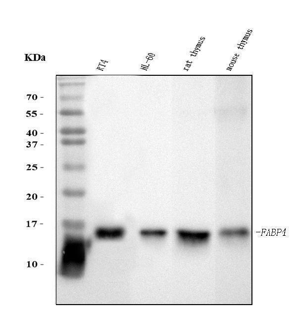
Figure 1. Western blot analysis of FABP4 using anti-FABP4 antibody (M01528-2).
Electrophoresis was performed on a 5-20% SDS-PAGE gel at 70V (Stacking gel) / 90V (Resolving gel) for 2-3 hours. The sample well of each lane was loaded with 30 ug of sample under reducing conditions.
Lane 1: human RT4 whole cell lysates,
Lane 2: human HL-60 whole cell lysates,
Lane 3: rat thymus tissue lysates,
Lane 4: mouse thymus tissue lysates.
After electrophoresis, proteins were transferred to a nitrocellulose membrane at 150 mA for 50-90 minutes. Blocked the membrane with 5% non-fat milk/TBS for 1.5 hour at RT. The membrane was incubated with mouse anti-FABP4 antigen affinity purified monoclonal antibody (Catalog # M01528-2) at 0.5 μg/mL overnight at 4°C, then washed with TBS-0.1%Tween 3 times with 5 minutes each and probed with a goat anti-mouse IgG-HRP secondary antibody at a dilution of 1:10000 for 1.5 hour at RT. The signal is developed using an Enhanced Chemiluminescent detection (ECL) kit (Catalog # EK1001) with Tanon 5200 system. A specific band was detected for FABP4 at approximately 15 kDa. The expected band size for FABP4 is at 15 kDa.

Click image to see more details
Figure 2. IHC analysis of FABP4 using anti-FABP4 antibody (M01528-2).
FABP4 was detected in a paraffin-embedded section of human renal clear cell carcinoma tissue. Heat mediated antigen retrieval was performed in EDTA buffer (pH 8.0, epitope retrieval solution). The tissue section was blocked with 10% goat serum. The tissue section was then incubated with 2 μg/ml mouse anti-FABP4 Antibody (M01528-2) overnight at 4°C. Biotinylated goat anti-mouse IgG was used as secondary antibody and incubated for 30 minutes at 37°C. The tissue section was developed using Strepavidin-Biotin-Complex (SABC) (Catalog # SA1021) with DAB as the chromogen.

Click image to see more details
Figure 3. IHC analysis of FABP4 using anti-FABP4 antibody (M01528-2).
FABP4 was detected in a paraffin-embedded section of human gall bladder adenosquamous carcinoma tissue. Heat mediated antigen retrieval was performed in EDTA buffer (pH 8.0, epitope retrieval solution). The tissue section was blocked with 10% goat serum. The tissue section was then incubated with 2 μg/ml mouse anti-FABP4 Antibody (M01528-2) overnight at 4°C. Biotinylated goat anti-mouse IgG was used as secondary antibody and incubated for 30 minutes at 37°C. The tissue section was developed using Strepavidin-Biotin-Complex (SABC) (Catalog # SA1021) with DAB as the chromogen.

Click image to see more details
Figure 4. IHC analysis of FABP4 using anti-FABP4 antibody (M01528-2).
FABP4 was detected in a paraffin-embedded section of human gastric cancer tissue. Heat mediated antigen retrieval was performed in EDTA buffer (pH 8.0, epitope retrieval solution). The tissue section was blocked with 10% goat serum. The tissue section was then incubated with 2 μg/ml mouse anti-FABP4 Antibody (M01528-2) overnight at 4°C. Biotinylated goat anti-mouse IgG was used as secondary antibody and incubated for 30 minutes at 37°C. The tissue section was developed using Strepavidin-Biotin-Complex (SABC) (Catalog # SA1021) with DAB as the chromogen.

Click image to see more details
Figure 5. IHC analysis of FABP4 using anti-FABP4 antibody (M01528-2).
FABP4 was detected in a paraffin-embedded section of human lymphadenoma tissue. Heat mediated antigen retrieval was performed in EDTA buffer (pH 8.0, epitope retrieval solution). The tissue section was blocked with 10% goat serum. The tissue section was then incubated with 2 μg/ml mouse anti-FABP4 Antibody (M01528-2) overnight at 4°C. Biotinylated goat anti-mouse IgG was used as secondary antibody and incubated for 30 minutes at 37°C. The tissue section was developed using Strepavidin-Biotin-Complex (SABC) (Catalog # SA1021) with DAB as the chromogen.

Click image to see more details
Figure 6. IHC analysis of FABP4 using anti-FABP4 antibody (M01528-2).
FABP4 was detected in a paraffin-embedded section of rat intestines tissue. Heat mediated antigen retrieval was performed in EDTA buffer (pH 8.0, epitope retrieval solution). The tissue section was blocked with 10% goat serum. The tissue section was then incubated with 2 μg/ml mouse anti-FABP4 Antibody (M01528-2) overnight at 4°C. Biotinylated goat anti-mouse IgG was used as secondary antibody and incubated for 30 minutes at 37°C. The tissue section was developed using Strepavidin-Biotin-Complex (SABC) (Catalog # SA1021) with DAB as the chromogen.

Click image to see more details
Figure 7. IHC analysis of FABP4 using anti-FABP4 antibody (M01528-2).
FABP4 was detected in a paraffin-embedded section of mouse intestines tissue. Heat mediated antigen retrieval was performed in EDTA buffer (pH 8.0, epitope retrieval solution). The tissue section was blocked with 10% goat serum. The tissue section was then incubated with 2 μg/ml mouse anti-FABP4 Antibody (M01528-2) overnight at 4°C. Biotinylated goat anti-mouse IgG was used as secondary antibody and incubated for 30 minutes at 37°C. The tissue section was developed using Strepavidin-Biotin-Complex (SABC) (Catalog # SA1021) with DAB as the chromogen.

Click image to see more details
Figure 8. IHC analysis of FABP4 using anti-FABP4 antibody (M01528-2).
FABP4 was detected in a paraffin-embedded section of mouse intestines tissue. Heat mediated antigen retrieval was performed in EDTA buffer (pH 8.0, epitope retrieval solution). The tissue section was blocked with 10% goat serum. The tissue section was then incubated with 2 μg/ml mouse anti-FABP4 Antibody (M01528-2) overnight at 4°C. Biotinylated goat anti-mouse IgG was used as secondary antibody and incubated for 30 minutes at 37°C. The tissue section was developed using Strepavidin-Biotin-Complex (SABC) (Catalog # SA1021) with DAB as the chromogen.
Protein Target Info & Infographic
Gene/Protein Information For FABP4 (Source: Uniprot.org, NCBI)
Gene Name
FABP4
Full Name
Fatty acid-binding protein, adipocyte
Weight
Superfamily
calycin superfamily
Alternative Names
Adipocyte lipid-binding protein; Adipocyte-type fatty acid-binding protein; AFABP; A-FABP; A-FABPAFABP; ALBP; aP2; FABP4; fatty acid binding protein 4, adipocyte; Fatty acid-binding protein 4; fatty acid-binding protein, adipocyte FABP4 A-FABP, AFABP, ALBP, HEL-S-104, aP2 fatty acid binding protein 4 fatty acid-binding protein, adipocyte|adipocyte fatty acid binding protein|adipocyte lipid-binding protein|adipocyte-type fatty acid-binding protein|epididymis secretory protein Li 104|fatty acid binding protein 4, adipocyte
*If product is indicated to react with multiple species, protein info is based on the gene entry specified above in "Species".For more info on FABP4, check out the FABP4 Infographic

We have 30,000+ of these available, one for each gene! Check them out.
In this infographic, you will see the following information for FABP4: database IDs, superfamily, protein function, synonyms, molecular weight, chromosomal locations, tissues of expression, subcellular locations, post-translational modifications, and related diseases, research areas & pathways. If you want to see more information included, or would like to contribute to it and be acknowledged, please contact [email protected].
Specific Publications For Anti-FABP4 Antibody Picoband™ (monoclonal, 10E12) (M01528-2)
Hello CJ!
No publications found for M01528-2
*Do you have publications using this product? Share with us and receive a reward. Ask us for more details.
Recommended Resources
Here are featured tools and databases that you might find useful.
- Boster's Pathways Library
- Protein Databases
- Bioscience Research Protocol Resources
- Data Processing & Analysis Software
- Photo Editing Software
- Scientific Literature Resources
- Research Paper Management Tools
- Molecular Biology Software
- Primer Design Tools
- Bioinformatics Tools
- Phylogenetic Tree Analysis
Customer Reviews
Have you used Anti-FABP4 Antibody Picoband™ (monoclonal, 10E12)?
Submit a review and receive an Amazon gift card.
- $30 for a review with an image
0 Reviews For Anti-FABP4 Antibody Picoband™ (monoclonal, 10E12)
Customer Q&As
Have a question?
Find answers in Q&As, reviews.
Can't find your answer?
Submit your question
