Click image to see more details
-

-
-
-
-
+2
Product Info Summary
| SKU: | M01357-4 |
|---|---|
| Size: | 100 μg/vial |
| Reactive Species: | Human |
| Host: | Mouse |
| Application: | Flow Cytometry, IF, IHC, IHC-F, ICC, WB |
Customers Who Bought This Also Bought

Product info
Product Name
Anti-Cytokeratin 18 KRT18 Antibody Picoband™(monoclonal, 7I6)
SKU/Catalog Number
M01357-4
Size
100 μg/vial
Form
Lyophilized
Description
Boster Bio Anti-Cytokeratin 18 KRT18 Antibody Picoband™ (monoclonal, 7I6) catalog # M01357-4. Tested in Flow Cytometry, IF, IHC, IHC-F, ICC, WB applications. This antibody reacts with Human.
Storage & Handling
Store at -20˚C for one year from date of receipt. After reconstitution, at 4˚C for one month. It can also be aliquotted and stored frozen at -20˚C for six months. Avoid repeated freeze-thaw cycles.
Cite This Product
Anti-Cytokeratin 18 KRT18 Antibody Picoband™(monoclonal, 7I6) (Boster Biological Technology, Pleasanton CA, USA, Catalog # M01357-4)
Host
Mouse
Contents
Each vial contains 4mg Trehalose, 0.9mg NaCl, 0.2mg Na2HPO4, 0.05mg NaN3.
Clonality
Monoclonal
Clone Number
7I6
Isotype
Mouse IgG1
Immunogen
E.coli-derived human Cytokeratin 18 recombinant protein (Position: E204-H430). Human Cytokeratin 18 shares 87.7% and 85.9% amino acid (aa) sequence identity with mouse and rat Cytokeratin 18, respectively.
*Blocking peptide can be purchased. Costs vary based on immunogen length. Contact us for pricing.
Cross-reactivity
No cross-reactivity with other proteins.
Reactive Species
M01357-4 is reactive to KRT18 in Human
Applications
M01357-4 is guaranteed for Flow Cytometry, IF, IHC, IHC-F, ICC, WB Boster Guarantee
Observed Molecular Weight
48 kDa
Calculated molecular weight
Background of KRT18
Keratin 18, mapped to 12q13.13, is a type I cytokeratin. It is, together with its filament partner keratin 8, perhaps the most commonly found products of the intermediate filament gene family. They are expressed in single layer epithelial tissues of the body. Mutations in this gene have been linked to cryptogenic cirrhosis. Two transcript variants encoding the same protein have been found for this gene.
Antibody Validation
Boster validates all antibodies on WB, IHC, ICC, Immunofluorescence, and ELISA with known positive control and negative samples to ensure specificity and high affinity, including thorough antibody incubations.
Assay dilution & Images
Reconstitution
Add 0.2ml of distilled water will yield a concentration of 500μg/ml.
Assay Dilutions Recommendation
The recommendations below provide a starting point for assay optimization. The actual working concentration varies and should be decided by the user.
Western blot, 0.1-0.5μg/ml, Human
Immunohistochemistry (Paraffin-embedded Section), 0.5-1μg/ml, Human
Immunohistochemistry (Frozen Section), 0.5-1μg/ml, Human
Immunocytochemistry/Immunofluorescence, 2μg/ml, Human
Immunofluorescence, 2μg/ml, Human
Flow Cytometry, 1-3μg/1x106 cells, Human
Validation Images & Assay Conditions
Click image to see more details
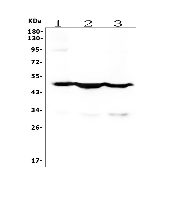
Figure 1. Western blot analysis of Cytokeratin 18 using anti-Cytokeratin 18 antibody (M01357-4).
Electrophoresis was performed on a 5-20% SDS-PAGE gel at 70V (Stacking gel) / 90V (Resolving gel) for 2-3 hours. The sample well of each lane was loaded with 50ug of sample under reducing conditions.
Lane 1: human placenta tissue lysates,
Lane 2: human Caco-2 whole cell lysates,
Lane 3: human A549 whole cell lysates.
After Electrophoresis, proteins were transferred to a Nitrocellulose membrane at 150mA for 50-90 minutes. Blocked the membrane with 5% Non-fat Milk/ TBS for 1.5 hours at RT. The membrane was incubated with mouse anti-Cytokeratin 18 antigen affinity purified monoclonal antibody (Catalog # M01357-4) at 0.5 μg/mL overnight at 4°C, then washed with TBS-0.1%Tween 3 times with 5 minutes each and probed with a goat anti-mouse IgG-HRP secondary antibody at a dilution of 1:10000 for 1.5 hour at RT. The signal is developed using an Enhanced Chemiluminescent detection (ECL) kit (Catalog # EK1001) with Tanon 5200 system. A specific band was detected for Cytokeratin 18 at approximately 48KD. The expected band size for Cytokeratin 18 is at 48KD.

Click image to see more details
Figure 2. IHC analysis of Cytokeratin 18 using anti-Cytokeratin 18 antibody (M01357-4).
Cytokeratin 18 was detected in paraffin-embedded section of human rectal cancer tissue. Heat mediated antigen retrieval was performed in EDTA buffer (pH8.0, epitope retrieval solution). The tissue section was blocked with 10% goat serum. The tissue section was then incubated with 1μg/ml mouse anti-Cytokeratin 18 Antibody (M01357-4) overnight at 4°C. Biotinylated goat anti-mouse IgG was used as secondary antibody and incubated for 30 minutes at 37°C. The tissue section was developed using Strepavidin-Biotin-Complex (SABC) (Catalog # SA1021) with DAB as the chromogen.

Click image to see more details
Figure 3. IHC analysis of Cytokeratin 18 using anti-Cytokeratin 18 antibody (M01357-4).
Cytokeratin 18 was detected in paraffin-embedded section of human lung cancer tissue. Heat mediated antigen retrieval was performed in EDTA buffer (pH8.0, epitope retrieval solution). The tissue section was blocked with 10% goat serum. The tissue section was then incubated with 1μg/ml mouse anti-Cytokeratin 18 Antibody (M01357-4) overnight at 4°C. Biotinylated goat anti-mouse IgG was used as secondary antibody and incubated for 30 minutes at 37°C. The tissue section was developed using Strepavidin-Biotin-Complex (SABC) (Catalog # SA1021) with DAB as the chromogen.

Click image to see more details
Figure 4. IF analysis of Cytokeratin 18 using anti-Cytokeratin 18 antibody (M01357-4).
Cytokeratin 18 was detected in immunocytochemical section of Hela cells. Enzyme antigen retrieval was performed using IHC enzyme antigen retrieval reagent (AR0022) for 15 mins. The cells were blocked with 10% goat serum. And then incubated with 2μg/mL mouse anti-Cytokeratin 18 Antibody (M01357-4) overnight at 4°C. DyLight®488 Conjugated Goat Anti-mouse IgG (BA1126) was used as secondary antibody at 1:100 dilution and incubated for 30 minutes at 37°C. The section was counterstained with DAPI. Visualize using a fluorescence microscope and filter sets appropriate for the label used.

Click image to see more details
Figure 5. IF analysis of Cytokeratin 18 using anti-Cytokeratin 18 antibody (M01357-4).
Cytokeratin 18 was detected in paraffin-embedded section of human placenta tissue. Heat mediated antigen retrieval was performed in EDTA buffer (pH8.0, epitope retrieval solution). The tissue section was blocked with 10% goat serum. The tissue section was then incubated with 1μg/mL mouse anti-Cytokeratin 18 Antibody (M01357-4) overnight at 4°C. DyLight®488 Conjugated Goat Anti-Mouse IgG (BA1126) was used as secondary antibody at 1:100 dilution and incubated for 30 minutes at 37°C. The section was counterstained with DAPI. Visualize using a fluorescence microscope and filter sets appropriate for the label used.

Click image to see more details
Figure 6. Flow Cytometry analysis of A431 cells using anti- Cytokeratin 18 antibody (M01357-4).
Overlay histogram showing A431 cells stained with M01357-4 (Blue line). To facilitate intracellular staining, cells were fixed with 4% paraformaldehyde and permeabilized with permeabilization buffer. The cells were blocked with 10% normal goat serum. And then incubated with mouse anti-Cytokeratin 18 Antibody (M01357-4, 1μg/1x106 cells) for 30 min at 20°C. DyLight®488 conjugated goat anti-mouse IgG (BA1126, 5-10μg/1x106 cells) was used as secondary antibody for 30 minutes at 20°C. Isotype control antibody (Green line) was mouse IgG (1μg/1x106) used under the same conditions. Unlabelled sample without incubation with primary antibody and secondary antibody (Red line) was used as a blank control.
Protein Target Info & Infographic
Gene/Protein Information For KRT18 (Source: Uniprot.org, NCBI)
Gene Name
KRT18
Full Name
Keratin, type I cytoskeletal 18
Weight
Superfamily
intermediate filament family
Alternative Names
Cell proliferation-inducing gene 46 protein; cell proliferation-inducing protein 46; CK-18; CYK18; Cytokeratin 18; cytokeratin-18; K18; Keratin 18; keratin, type I cytoskeletal 18; keratin-18; KRT18 KRT18 CK-18, CYK18, K18 keratin 18 keratin, type I cytoskeletal 18|cell proliferation-inducing gene 46 protein|cytokeratin 18|keratin 18, type I
*If product is indicated to react with multiple species, protein info is based on the gene entry specified above in "Species".For more info on KRT18, check out the KRT18 Infographic

We have 30,000+ of these available, one for each gene! Check them out.
In this infographic, you will see the following information for KRT18: database IDs, superfamily, protein function, synonyms, molecular weight, chromosomal locations, tissues of expression, subcellular locations, post-translational modifications, and related diseases, research areas & pathways. If you want to see more information included, or would like to contribute to it and be acknowledged, please contact [email protected].
Specific Publications For Anti-Cytokeratin 18 KRT18 Antibody Picoband™(monoclonal, 7I6) (M01357-4)
Hello CJ!
M01357-4 has been cited in 2 publications:
*The publications in this section are manually curated by our staff scientists. They may differ from Bioz's machine gathered results. Both are accurate. If you find a publication citing this product but is missing from this list, please let us know we will issue you a thank-you coupon.
Wei Z,Wang J,Wang Y,Wang C,Liu X,Han Z,Fu Y,Yang Z.Effects of Neutrophil Extracellular Traps on Bovine Mammary Epithelial Cells in vitro.Front Immunol.2019 May 17;10:1003.doi:10.3389/fimmu.2019.01003.PMID:31156617;PMCID:PMC6533846.
Species: Cows
M01357-4 usage in article: APP:IF, SAMPLE:BMECS, DILUTION: NA
Shi R,Liu L,Wang F,He Y,Niu Y,Wang C,Zhang X,Zhang X,Zhang H,Chen M,Wang Y.Downregulation of cytokeratin 18 induces cellular partial EMT and stemness through increasing EpCAM expression in breast cancer.Cell Signal.2020 Dec;76:109810.doi:10.1016/j.cellsig
Species: Human
M01357-4 usage in article: APP:IF, SAMPLE:BREAST CANCER CELLS AND BREAST TISSUE, DILUTION:1:100
Recommended Resources
Here are featured tools and databases that you might find useful.
- Boster's Pathways Library
- Protein Databases
- Bioscience Research Protocol Resources
- Data Processing & Analysis Software
- Photo Editing Software
- Scientific Literature Resources
- Research Paper Management Tools
- Molecular Biology Software
- Primer Design Tools
- Bioinformatics Tools
- Phylogenetic Tree Analysis
Customer Reviews
Have you used Anti-Cytokeratin 18 KRT18 Antibody Picoband™(monoclonal, 7I6)?
Submit a review and receive an Amazon gift card.
- $30 for a review with an image
0 Reviews For Anti-Cytokeratin 18 KRT18 Antibody Picoband™(monoclonal, 7I6)
Customer Q&As
Have a question?
Find answers in Q&As, reviews.
Can't find your answer?
Submit your question
