Click image to see more details
-

-
-
-
-
+10
Product Info Summary
| SKU: | A04722-1 |
|---|---|
| Size: | 100 μg/vial |
| Reactive Species: | Human, Mouse, Rat |
| Host: | Rabbit |
| Application: | Flow Cytometry, IF, ICC, WB |
Customers Who Bought This Also Bought

Product info
Product Name
Anti-CSNK2A2 Antibody Picoband®
View all CKII alpha prime polypeptide Antibodies
SKU/Catalog Number
A04722-1
Size
100 μg/vial
Form
Lyophilized
Description
Boster Bio Anti-CSNK2A2 Antibody Picoband® catalog # A04722-1. Tested in Flow Cytometry, IF, ICC, WB applications. This antibody reacts with Human, Mouse, Rat. The brand Picoband indicates this is a premium antibody that guarantees superior quality, high affinity, and strong signals with minimal background in Western blot applications. Only our best-performing antibodies are designated as Picoband, ensuring unmatched performance.
Storage & Handling
At -20°C for one year from date of receipt. After reconstitution, at 4°C for one month. It can also be aliquotted and stored frozen at -20°C for six months. Avoid repeated freezing and thawing.
Cite This Product
Anti-CSNK2A2 Antibody Picoband® (Boster Biological Technology, Pleasanton CA, USA, Catalog # A04722-1)
Host
Rabbit
Contents
Each vial contains 4 mg Trehalose, 0.9 mg NaCl, 0.2 mg Na2HPO4.
Clonality
Polyclonal
Isotype
Rabbit IgG
Immunogen
A synthetic peptide corresponding to a sequence at the C-terminus of human CSNK2A2, which shares 93.3% amino acid (aa) sequence identity with mouse CSNK2A2.
*Blocking peptide can be purchased. Costs vary based on immunogen length. Contact us for pricing.
Cross-reactivity
No cross-reactivity with other proteins.
Reactive Species
A04722-1 is reactive to CSNK2A2 in Human, Mouse, Rat
Reconstitution
Adding 0.2 ml of distilled water will yield a concentration of 500 μg/ml.
Observed Molecular Weight
39 kDa
Calculated molecular weight
41.213kDa
Background of CKII alpha prime polypeptide
Casein kinase II subunit alpha' is an enzyme that in humans is encoded by the CSNK2A2 gene. This gene encodes the alpha', or alpha 2, catalytic subunit of the protein kinase enzyme, casein kinase 2 (CK2). Casein kinase 2 is a serine/threonine protein kinase that phosphorylates acidic proteins such as casein. It is involved in various cellular processes, including cell cycle control, apoptosis, and circadian rhythms. This heterotetrameric kinase includes two catalytic subunits, either alpha or alpha', and two regulatory beta subunits. The closely related gene paralog encoding the alpha, or alpha 1 subunit (CSNK2A1, Gene ID: 1457) is found on chromosome 20. An intronic variant in this gene (alpha 2) may be associated with leukocyte telomere length in a South Asian population. A related transcribed pseudogene is found on chromosome 11.
Antibody Validation
Boster validates all antibodies on WB, IHC, ICC, Immunofluorescence, and ELISA with known positive control and negative samples to ensure specificity and high affinity, including thorough antibody incubations.
Application & Images
Applications
A04722-1 is guaranteed for Flow Cytometry, IF, ICC, WB Boster Guarantee
Assay Dilutions Recommendation
The recommendations below provide a starting point for assay optimization. The actual working concentration varies and should be decided by the user.
Western blot, 0.25-0.5 μg/ml, Human, Mouse, Rat
Immunocytochemistry/Immunofluorescence, 5 μg/ml, Human
Flow Cytometry (Fixed), 1-3 μg/1x106 cells, Human
Positive Control
WB: human Hela whole cell, human 293T whole cell, human SiHa whole cell, human THP-1 whole cell, rat testis tissue, rat thymus tissue, mouse testis tissue, mouse thymus tissue
IHC: human bladder epithelial carcinoma tissue, human endometrial cancer tissue, human laryngeal squamous cell carcinoma tissue, human colonic adenocarcinoma tissue, human placenta tissue, human liver cancer tissue, mouse colon tissue, mouse testis tissue, rat colon tissue, rat testis tissue
ICC/IF: Caco-2 cell
FCM: Caco-2 cell, THP-1 cell
Validation Images & Assay Conditions
Click image to see more details
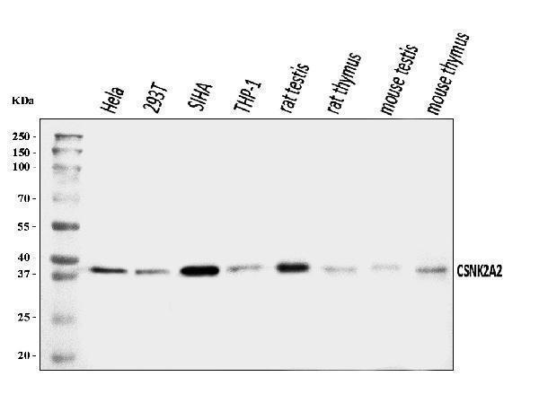
Figure 1. Western blot analysis of CSNK2A2 using anti-CSNK2A2 antibody (A04722-1).
Electrophoresis was performed on a 5-20% SDS-PAGE gel at 70V (Stacking gel) / 90V (Resolving gel) for 2-3 hours. The sample well of each lane was loaded with 30 ug of sample under reducing conditions.
Lane 1: human Hela whole cell lysates,
Lane 2: human 293T whole cell lysates,
Lane 3: human SiHa whole cell lysates,
Lane 4: human THP-1 whole cell lysates,
Lane 5: rat testis tissue lysates,
Lane 6: rat thymus tissue lysates,
Lane 7: mouse testis tissue lysates,
Lane 8: mouse thymus tissue lysates.
After electrophoresis, proteins were transferred to a nitrocellulose membrane at 150 mA for 50-90 minutes. Blocked the membrane with 5% non-fat milk/TBS for 1.5 hour at RT. The membrane was incubated with rabbit anti-CSNK2A2 antigen affinity purified polyclonal antibody (Catalog # A04722-1) at 0.5 μg/mL overnight at 4°C, then washed with TBS-0.1%Tween 3 times with 5 minutes each and probed with a goat anti-rabbit IgG-HRP secondary antibody at a dilution of 1:5000 for 1.5 hour at RT. The signal is developed using an Enhanced Chemiluminescent detection (ECL) kit (Catalog # EK1002) with Tanon 5200 system. A specific band was detected for CSNK2A2 at approximately 39 kDa. The expected band size for CSNK2A2 is at 41 kDa.

Click image to see more details
Figure 2. IHC analysis of CSNK2K2 using anti-CSNK2K2 antibody (A04722-1).
CSNK2K2 was detected in a paraffin-embedded section of human bladder epithelial carcinoma tissue. Heat mediated antigen retrieval was performed in EDTA buffer (pH 8.0, epitope retrieval solution). The tissue section was blocked with 10% goat serum. The tissue section was then incubated with 2 μg/ml rabbit anti-CSNK2K2 Antibody (A04722-1) overnight at 4°C. Biotinylated goat anti-rabbit IgG was used as secondary antibody and incubated for 30 minutes at 37°C. The tissue section was developed using Strepavidin-Biotin-Complex (SABC) (Catalog # SA1022) with DAB as the chromogen.

Click image to see more details
Figure 3. IHC analysis of CSNK2K2 using anti-CSNK2K2 antibody (A04722-1).
CSNK2K2 was detected in a paraffin-embedded section of human endometrial cancer tissue. Heat mediated antigen retrieval was performed in EDTA buffer (pH 8.0, epitope retrieval solution). The tissue section was blocked with 10% goat serum. The tissue section was then incubated with 2 μg/ml rabbit anti-CSNK2K2 Antibody (A04722-1) overnight at 4°C. Biotinylated goat anti-rabbit IgG was used as secondary antibody and incubated for 30 minutes at 37°C. The tissue section was developed using Strepavidin-Biotin-Complex (SABC) (Catalog # SA1022) with DAB as the chromogen.

Click image to see more details
Figure 4. IHC analysis of CSNK2K2 using anti-CSNK2K2 antibody (A04722-1).
CSNK2K2 was detected in a paraffin-embedded section of human laryngeal squamous cell carcinoma tissue. Heat mediated antigen retrieval was performed in EDTA buffer (pH 8.0, epitope retrieval solution). The tissue section was blocked with 10% goat serum. The tissue section was then incubated with 2 μg/ml rabbit anti-CSNK2K2 Antibody (A04722-1) overnight at 4°C. Biotinylated goat anti-rabbit IgG was used as secondary antibody and incubated for 30 minutes at 37°C. The tissue section was developed using Strepavidin-Biotin-Complex (SABC) (Catalog # SA1022) with DAB as the chromogen.

Click image to see more details
Figure 5. IHC analysis of CSNK2K2 using anti-CSNK2K2 antibody (A04722-1).
CSNK2K2 was detected in a paraffin-embedded section of human colonic adenocarcinoma tissue. Heat mediated antigen retrieval was performed in EDTA buffer (pH 8.0, epitope retrieval solution). The tissue section was blocked with 10% goat serum. The tissue section was then incubated with 2 μg/ml rabbit anti-CSNK2K2 Antibody (A04722-1) overnight at 4°C. Biotinylated goat anti-rabbit IgG was used as secondary antibody and incubated for 30 minutes at 37°C. The tissue section was developed using Strepavidin-Biotin-Complex (SABC) (Catalog # SA1022) with DAB as the chromogen.

Click image to see more details
Figure 6. IHC analysis of CSNK2K2 using anti-CSNK2K2 antibody (A04722-1).
CSNK2K2 was detected in a paraffin-embedded section of human placenta tissue. Heat mediated antigen retrieval was performed in EDTA buffer (pH 8.0, epitope retrieval solution). The tissue section was blocked with 10% goat serum. The tissue section was then incubated with 2 μg/ml rabbit anti-CSNK2K2 Antibody (A04722-1) overnight at 4°C. Biotinylated goat anti-rabbit IgG was used as secondary antibody and incubated for 30 minutes at 37°C. The tissue section was developed using Strepavidin-Biotin-Complex (SABC) (Catalog # SA1022) with DAB as the chromogen.

Click image to see more details
Figure 7. IHC analysis of CSNK2K2 using anti-CSNK2K2 antibody (A04722-1).
CSNK2K2 was detected in a paraffin-embedded section of human liver cancer tissue. Heat mediated antigen retrieval was performed in EDTA buffer (pH 8.0, epitope retrieval solution). The tissue section was blocked with 10% goat serum. The tissue section was then incubated with 2 μg/ml rabbit anti-CSNK2K2 Antibody (A04722-1) overnight at 4°C. Biotinylated goat anti-rabbit IgG was used as secondary antibody and incubated for 30 minutes at 37°C. The tissue section was developed using Strepavidin-Biotin-Complex (SABC) (Catalog # SA1022) with DAB as the chromogen.

Click image to see more details
Figure 8. IHC analysis of CSNK2K2 using anti-CSNK2K2 antibody (A04722-1).
CSNK2K2 was detected in a paraffin-embedded section of mouse colon tissue. Heat mediated antigen retrieval was performed in EDTA buffer (pH 8.0, epitope retrieval solution). The tissue section was blocked with 10% goat serum. The tissue section was then incubated with 2 μg/ml rabbit anti-CSNK2K2 Antibody (A04722-1) overnight at 4°C. Biotinylated goat anti-rabbit IgG was used as secondary antibody and incubated for 30 minutes at 37°C. The tissue section was developed using Strepavidin-Biotin-Complex (SABC) (Catalog # SA1022) with DAB as the chromogen.

Click image to see more details
Figure 13. Flow Cytometry analysis of Caco-2 cells using anti-CSNK2A2 antibody (A04722-1).
Overlay histogram showing Caco-2 cells stained with A04722-1 (Blue line). To facilitate intracellular staining, cells were fixed with 4% paraformaldehyde and permeabilized with permeabilization buffer. The cells were blocked with 10% normal goat serum. And then incubated with rabbit anti-CSNK2A2 Antibody (A04722-1, 1 μg/1x106 cells) for 30 min at 20°C. DyLight®488 conjugated goat anti-rabbit IgG (BA1127, 5-10 μg/1x106 cells) was used as secondary antibody for 30 minutes at 20°C. Isotype control antibody (Green line) was rabbit IgG (1 μg/1x106) used under the same conditions. Unlabelled sample without incubation with primary antibody and secondary antibody (Red line) was used as a blank control.

Click image to see more details
Figure 9. IHC analysis of CSNK2K2 using anti-CSNK2K2 antibody (A04722-1).
CSNK2K2 was detected in a paraffin-embedded section of mouse testis tissue. Heat mediated antigen retrieval was performed in EDTA buffer (pH 8.0, epitope retrieval solution). The tissue section was blocked with 10% goat serum. The tissue section was then incubated with 2 μg/ml rabbit anti-CSNK2K2 Antibody (A04722-1) overnight at 4°C. Biotinylated goat anti-rabbit IgG was used as secondary antibody and incubated for 30 minutes at 37°C. The tissue section was developed using Strepavidin-Biotin-Complex (SABC) (Catalog # SA1022) with DAB as the chromogen.

Click image to see more details
Figure 14. Flow Cytometry analysis of THP-1 cells using anti-CSNK2A2 antibody (A04722-1).
Overlay histogram showing THP-1 cells stained with A04722-1 (Blue line). To facilitate intracellular staining, cells were fixed with 4% paraformaldehyde and permeabilized with permeabilization buffer. The cells were blocked with 10% normal goat serum. And then incubated with rabbit anti-CSNK2A2 Antibody (A04722-1, 1 μg/1x106 cells) for 30 min at 20°C. DyLight®488 conjugated goat anti-rabbit IgG (BA1127, 5-10 μg/1x106 cells) was used as secondary antibody for 30 minutes at 20°C. Isotype control antibody (Green line) was rabbit IgG (1 μg/1x106) used under the same conditions. Unlabelled sample without incubation with primary antibody and secondary antibody (Red line) was used as a blank control.

Click image to see more details
Figure 10. IHC analysis of CSNK2K2 using anti-CSNK2K2 antibody (A04722-1).
CSNK2K2 was detected in a paraffin-embedded section of rat colon tissue. Heat mediated antigen retrieval was performed in EDTA buffer (pH 8.0, epitope retrieval solution). The tissue section was blocked with 10% goat serum. The tissue section was then incubated with 2 μg/ml rabbit anti-CSNK2K2 Antibody (A04722-1) overnight at 4°C. Biotinylated goat anti-rabbit IgG was used as secondary antibody and incubated for 30 minutes at 37°C. The tissue section was developed using Strepavidin-Biotin-Complex (SABC) (Catalog # SA1022) with DAB as the chromogen.

Click image to see more details
Figure 11. IHC analysis of CSNK2K2 using anti-CSNK2K2 antibody (A04722-1).
CSNK2K2 was detected in a paraffin-embedded section of rat testis tissue. Heat mediated antigen retrieval was performed in EDTA buffer (pH 8.0, epitope retrieval solution). The tissue section was blocked with 10% goat serum. The tissue section was then incubated with 2 μg/ml rabbit anti-CSNK2K2 Antibody (A04722-1) overnight at 4°C. Biotinylated goat anti-rabbit IgG was used as secondary antibody and incubated for 30 minutes at 37°C. The tissue section was developed using Strepavidin-Biotin-Complex (SABC) (Catalog # SA1022) with DAB as the chromogen.

Click image to see more details
Figure 12. IF analysis of CSNK2A2 using anti-CSNK2A2 antibody (A04722-1).
CSNK2A2 was detected in an immunocytochemical section of Caco-2 cells. Enzyme antigen retrieval was performed using IHC enzyme antigen retrieval reagent (AR0022) for 15 mins. The cells were blocked with 10% goat serum. And then incubated with 5 μg/mL rabbit anti-CSNK2A2 Antibody (A04722-1) overnight at 4°C. DyLight®488 Conjugated Goat Anti-Rabbit IgG (BA1127) was used as secondary antibody at 1:100 dilution and incubated for 30 minutes at 37°C. The section was counterstained with DAPI. Visualize using a fluorescence microscope and filter sets appropriate for the label used.
Protein Target Info & Infographic
Gene/Protein Information For CSNK2A2 (Source: Uniprot.org, NCBI)
Gene Name
CSNK2A2
Full Name
Casein kinase II subunit alpha'
Weight
41.213kDa
Superfamily
protein kinase superfamily
Alternative Names
Heat shock factor protein 2; HSF 2; Heat shock transcription factor 2; HSTF 2; HSF2; HSTF2 CSNK2A2 CK2A2, CK2alpha, CSNK2A1 casein kinase 2 alpha 2 casein kinase II subunit alpha|CK II alpha|casein kinase 2 alpha|casein kinase 2, alpha prime polypeptide
*If product is indicated to react with multiple species, protein info is based on the gene entry specified above in "Species".For more info on CSNK2A2, check out the CSNK2A2 Infographic

We have 30,000+ of these available, one for each gene! Check them out.
In this infographic, you will see the following information for CSNK2A2: database IDs, superfamily, protein function, synonyms, molecular weight, chromosomal locations, tissues of expression, subcellular locations, post-translational modifications, and related diseases, research areas & pathways. If you want to see more information included, or would like to contribute to it and be acknowledged, please contact [email protected].
Specific Publications For Anti-CSNK2A2 Antibody Picoband® (A04722-1)
Hello CJ!
No publications found for A04722-1
*Do you have publications using this product? Share with us and receive a reward. Ask us for more details.
Recommended Resources
Here are featured tools and databases that you might find useful.
- Boster's Pathways Library
- Protein Databases
- Bioscience Research Protocol Resources
- Data Processing & Analysis Software
- Photo Editing Software
- Scientific Literature Resources
- Research Paper Management Tools
- Molecular Biology Software
- Primer Design Tools
- Bioinformatics Tools
- Phylogenetic Tree Analysis
Customer Reviews
Have you used Anti-CSNK2A2 Antibody Picoband®?
Submit a review and receive an Amazon gift card.
- $30 for a review with an image
0 Reviews For Anti-CSNK2A2 Antibody Picoband®
Customer Q&As
Have a question?
Find answers in Q&As, reviews.
Can't find your answer?
Submit your question
