Click image to see more details
Product Info Summary
| SKU: | A01094-3 |
|---|---|
| Size: | 100 μg/vial |
| Reactive Species: | Human, Mouse |
| Host: | Rabbit |
| Application: | ELISA, Flow Cytometry, WB |
Customers Who Bought This Also Bought

Product info
Product Name
Anti-CD226 Antibody Picoband™
View all DNAM-1/CD226 Antibodies
SKU/Catalog Number
A01094-3
Size
100 μg/vial
Form
Lyophilized
Description
Boster Bio Anti-CD226 Antibody Picoband™ catalog # A01094-3. Tested in ELISA, Flow Cytometry, WB applications. This antibody reacts with Human, Mouse.
Storage & Handling
At -20°C for one year from date of receipt. After reconstitution, at 4°C for one month. It can also be aliquotted and stored frozen at -20°C for six months. Avoid repeated freezing and thawing.
Cite This Product
Anti-CD226 Antibody Picoband™ (Boster Biological Technology, Pleasanton CA, USA, Catalog # A01094-3)
Host
Rabbit
Contents
Each vial contains 4 mg Trehalose, 0.9 mg NaCl, 0.2 mg Na2HPO4.
Clonality
Polyclonal
Isotype
Rabbit IgG
Immunogen
E.coli-derived human CD226 recombinant protein (Position: E19-A254).
*Blocking peptide can be purchased. Costs vary based on immunogen length. Contact us for pricing.
Cross-reactivity
No cross-reactivity with other proteins
Reactive Species
A01094-3 is reactive to CD226 in Human, Mouse
Applications
A01094-3 is guaranteed for ELISA, Flow Cytometry, WB Boster Guarantee
Observed Molecular Weight
70 kDa
Calculated molecular weight
122762 MW
Background of DNAM-1/CD226
DNAM 1, also known as CD226, is a receptor expressed by peripheral blood T lymphocytes that is involved in intercellular adhesion, lymphocyte signaling, cytotoxicity and lymphokine secretion mediated by cytotoxic T lymphocytes. DNAM 1 is broadly expressed on T cells, NK cells, platelets, monocytes and a subset of B cells. DNAM-1 is also expressed by a subset of CD3 positive thymocytes. This antibody is reported to inhibit T- and NK cell mediated cytotoxicity against tumour cell targets and to block TNF alpha and IFN gamma secretion by alloantigen-specific T-cells.
Antibody Validation
Boster validates all antibodies on WB, IHC, ICC, Immunofluorescence, and ELISA with known positive control and negative samples to ensure specificity and high affinity, including thorough antibody incubations.
Assay dilution & Images
Reconstitution
Adding 0.2 ml of distilled water will yield a concentration of 500 μg/ml.
Assay Dilutions Recommendation
The recommendations below provide a starting point for assay optimization. The actual working concentration varies and should be decided by the user.
Western blot, 0.25-0.5 μg/ml, Human, Mouse
Flow Cytometry (Fixed), 1-3 μg/1x106 cells, Human
Direct ELISA, 0.1-0.5 μg/ml, Human
Validation Images & Assay Conditions
Click image to see more details
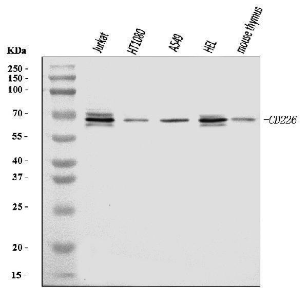
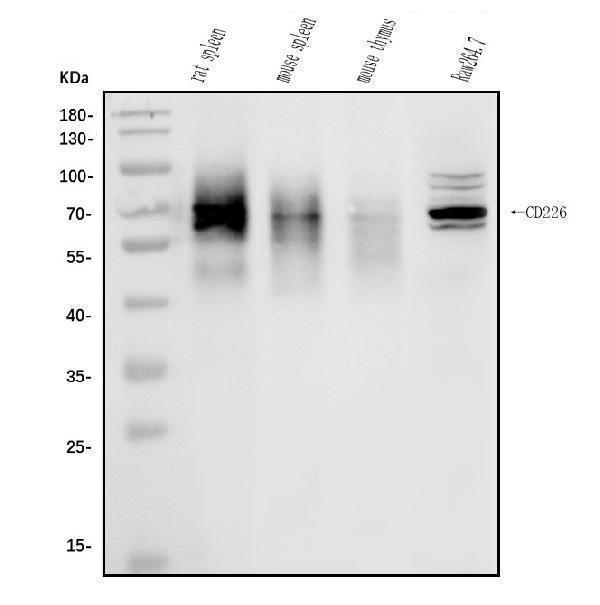
Figure 1. Western blot analysis of CD226 using anti-CD226 antibody (A01094-3).
Electrophoresis was performed on a 5-20% SDS-PAGE gel at 70V (Stacking gel) / 90V (Resolving gel) for 2-3 hours. The sample well of each lane was loaded with 30 ug of sample under reducing conditions.
Lane 1: human Jurkat whole cell lysates,
Lane 2: human HT1080 whole cell lysates,
Lane 3: human A549 whole cell lysates,
Lane 4: human HEL whole cell lysates,
Lane 5: mouse thymus tissue lysates.
After electrophoresis, proteins were transferred to a nitrocellulose membrane at 150 mA for 50-90 minutes. Blocked the membrane with 5% non-fat milk/TBS for 1.5 hour at RT. The membrane was incubated with rabbit anti-CD226 antigen affinity purified polyclonal antibody (Catalog # A01094-3) at 0.5 μg/mL overnight at 4°C, then washed with TBS-0.1%Tween 3 times with 5 minutes each and probed with a goat anti-rabbit IgG-HRP secondary antibody at a dilution of 1:5000 for 1.5 hour at RT. The signal is developed using an Enhanced Chemiluminescent detection (ECL) kit (Catalog # EK1002) with Tanon 5200 system. A specific band was detected for CD226 at approximately 70 kDa. The expected band size for CD226 is at 70 kDa.

Click image to see more details
Figure 2. Flow Cytometry analysis of Jurkat cells using anti-CD226 antibody (A01094-3).
Overlay histogram showing Jurkat cells stained with A01094-3 (Blue line). To facilitate intracellular staining, cells were fixed with 4% paraformaldehyde and permeabilized with permeabilization buffer. The cells were blocked with 10% normal goat serum. And then incubated with rabbit anti-CD226 Antibody (A01094-3, 1 μg/1x106 cells) for 30 min at 20°C. DyLight®488 conjugated goat anti-rabbit IgG (BA1127, 5-10 μg/1x106 cells) was used as secondary antibody for 30 minutes at 20°C. Isotype control antibody (Green line) was rabbit IgG (1 μg/1x106) used under the same conditions. Unlabelled sample without incubation with primary antibody and secondary antibody (Red line) was used as a blank control.
Protein Target Info & Infographic
Gene/Protein Information For CD226 (Source: Uniprot.org, NCBI)
Gene Name
CD226
Full Name
CD226 antigen
Weight
122762 MW
Alternative Names
CD226 antigenplatelet and T cell activation antigen 1; CD226 molecule; CD226; DNAM1; DNAM-1; DNAM1adhesion glycoprotein; DNAM-1DNAX accessory molecule-1; DNAX accessory molecule 1; PTA1; T lineage-specific activation antigen 1 antigen; TLiSA1 CD226 DNAM-1, DNAM1, PTA1, TLiSA1 CD226 molecule CD226 antigen|DNAX accessory molecule-1|T lineage-specific activation antigen 1 antigen|adhesion glycoprotein|platelet and T cell activation antigen 1
*If product is indicated to react with multiple species, protein info is based on the gene entry specified above in "Species".For more info on CD226, check out the CD226 Infographic

We have 30,000+ of these available, one for each gene! Check them out.
In this infographic, you will see the following information for CD226: database IDs, superfamily, protein function, synonyms, molecular weight, chromosomal locations, tissues of expression, subcellular locations, post-translational modifications, and related diseases, research areas & pathways. If you want to see more information included, or would like to contribute to it and be acknowledged, please contact [email protected].
Specific Publications For Anti-CD226 Antibody Picoband™ (A01094-3)
Hello CJ!
No publications found for A01094-3
*Do you have publications using this product? Share with us and receive a reward. Ask us for more details.
Recommended Resources
Here are featured tools and databases that you might find useful.
- Boster's Pathways Library
- Protein Databases
- Bioscience Research Protocol Resources
- Data Processing & Analysis Software
- Photo Editing Software
- Scientific Literature Resources
- Research Paper Management Tools
- Molecular Biology Software
- Primer Design Tools
- Bioinformatics Tools
- Phylogenetic Tree Analysis
Customer Reviews
Have you used Anti-CD226 Antibody Picoband™?
Submit a review and receive an Amazon gift card.
- $30 for a review with an image
0 Reviews For Anti-CD226 Antibody Picoband™
Customer Q&As
Have a question?
Find answers in Q&As, reviews.
Can't find your answer?
Submit your question
