
Click image to see more details
-

-
-
-
-
+2
Product Info Summary
| SKU: | PB9977 |
|---|---|
| Size: | 100 μg/vial |
| Reactive Species: | Human |
| Host: | Rabbit |
| Application: | Flow Cytometry, IF, IHC, ICC, WB |
Customers Who Bought This Also Bought

Product info
Product Name
Anti-AEBP2 Antibody Picoband®
SKU/Catalog Number
PB9977
PB1029 is an alternative SKU for this antibody, used in previous lots.
Size
100 μg/vial
Form
Lyophilized
Description
Boster Bio Anti-AEBP2 Antibody Picoband® catalog # PB9977. Tested in Flow Cytometry, IF, IHC, ICC, WB applications. This antibody reacts with Human. The brand Picoband indicates this is a premium antibody that guarantees superior quality, high affinity, and strong signals with minimal background in Western blot applications. Only our best-performing antibodies are designated as Picoband, ensuring unmatched performance.
Storage & Handling
Store at -20˚C for one year from date of receipt. After reconstitution, at 4˚C for one month. It can also be aliquotted and stored frozen at -20˚C for six months. Avoid repeated freeze-thaw cycles.
Cite This Product
Anti-AEBP2 Antibody Picoband® (Boster Biological Technology, Pleasanton CA, USA, Catalog # PB9977)
Host
Rabbit
Contents
Each vial contains 5mg BSA, 0.9mg NaCl, 0.2mg Na2HPO4, 0.05mg NaN3.
Clonality
Polyclonal
Isotype
Rabbit IgG
Immunogen
E.coli-derived human AEBP2 recombinant protein (Position: K424-Q517). Human AEBP2 shares 98.8% amino acid (aa) sequence identity with mouse AEBP2.
*Blocking peptide can be purchased. Costs vary based on immunogen length. Contact us for pricing.
Cross-reactivity
No cross-reactivity with other proteins.
Reactive Species
PB9977 is reactive to AEBP2 in Human
Reconstitution
Add 0.2ml of distilled water will yield a concentration of 500ug/ml.
Observed Molecular Weight
54 kDa
Calculated molecular weight
54467MW
Background of Aebp2
Adipocyte Enhancer-Binding Protein is a zinc finger protein that in humans is encoded by the evolutionarily well-conserved gene AEBP2. This gene is mapped to 12p12.3. AEBP2 is a DNA-binding transcriptional repressor. It may regulate the migration and development of the neural crest cells through the PRC2-mediated epigenetic mechanism and is most likely a targeting protein for the mammalian PRC2 complex.
Antibody Validation
Boster validates all antibodies on WB, IHC, ICC, Immunofluorescence, and ELISA with known positive control and negative samples to ensure specificity and high affinity, including thorough antibody incubations.
Application & Images
Applications
PB9977 is guaranteed for Flow Cytometry, IF, IHC, ICC, WB Boster Guarantee
Assay Dilutions Recommendation
The recommendations below provide a starting point for assay optimization. The actual working concentration varies and should be decided by the user.
Western blot, 0.1-0.5μg/ml, Human
Immunohistochemistry (Paraffin-embedded Section), 0.5-1μg/ml, Human, By Heat
Immunocytochemistry/Immunofluorescence, 2μg/ml, Human
Flow Cytometry (Fixed), 1-3μg/1x106 cells, Human
Positive Control
WB: human Hela whole cell, human Jurkat whole cell, human K562 whole cell, human HL-60 whole cell, human RT4 whole cell, human SiHa whole cell, human CACO-2 whole cell, human A549 whole cell
IHC: human lung cancer tissue, human mammary cancer tissue, human placenta tissue
ICC/IF: U20S cell
FCM: SiHa cell
Validation Images & Assay Conditions
Click image to see more details
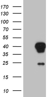
Figure 1. Western blot analysis of AEBP2 using anti-AEBP2 antibody (PB9977).
Electrophoresis was performed on a 5-20% SDS-PAGE gel at 70V (Stacking gel) / 90V (Resolving gel) for 2-3 hours. The sample well of each lane was loaded with 30 ug of sample under reducing conditions.
Lane 1: human Hela whole cell lysates,
Lane 2: human Jurkat whole cell lysates,
Lane 3: human K562 whole cell lysates,
Lane 4: human HL-60 whole cell lysates,
Lane 5: human RT4 whole cell lysates,
Lane 6: human SiHa whole cell lysates,
Lane 7: human CACO-2 whole cell lysates,
Lane 8: human A549 whole cell lysates.
After electrophoresis, proteins were transferred to a nitrocellulose membrane at 150 mA for 50-90 minutes. Blocked the membrane with 5% non-fat milk/TBS for 1.5 hour at RT. The membrane was incubated with rabbit anti-AEBP2 antigen affinity purified polyclonal antibody (Catalog # PB9977) at 0.5 μg/mL overnight at 4°C, then washed with TBS-0.1%Tween 3 times with 5 minutes each and probed with a goat anti-rabbit IgG-HRP secondary antibody at a dilution of 1:5000 for 1.5 hour at RT. The signal is developed using an Enhanced Chemiluminescent detection (ECL) kit (Catalog # EK1002) with Tanon 5200 system. A specific band was detected for AEBP2 at approximately 54 kDa. The expected band size for AEBP2 is at 54 kDa.

Click image to see more details
Figure 2. IHC analysis of AEBP2 using anti-AEBP2 antibody (PB9977).
AEBP2 was detected in paraffin-embedded section of human lung cancer tissues. Heat mediated antigen retrieval was performed in citrate buffer (pH6, epitope retrieval solution) for 20 mins. The tissue section was blocked with 10% goat serum. The tissue section was then incubated with 1μg/ml rabbit anti-AEBP2 Antibody (PB9977) overnight at 4°C. Biotinylated goat anti-rabbit IgG was used as secondary antibody and incubated for 30 minutes at 37°C. The tissue section was developed using Strepavidin-Biotin-Complex (SABC)(Catalog # SA1022) with DAB as the chromogen.

Click image to see more details
Figure 3. IHC analysis of AEBP2 using anti-AEBP2 antibody (PB9977).
AEBP2 was detected in paraffin-embedded section of human mammary cancer tissues. Heat mediated antigen retrieval was performed in citrate buffer (pH6, epitope retrieval solution) for 20 mins. The tissue section was blocked with 10% goat serum. The tissue section was then incubated with 1μg/ml rabbit anti-AEBP2 Antibody (PB9977) overnight at 4°C. Biotinylated goat anti-rabbit IgG was used as secondary antibody and incubated for 30 minutes at 37°C. The tissue section was developed using Strepavidin-Biotin-Complex (SABC)(Catalog # SA1022) with DAB as the chromogen.

Click image to see more details
Figure 4. IHC analysis of AEBP2 using anti-AEBP2 antibody (PB9977).
AEBP2 was detected in paraffin-embedded section of human placenta tissues. Heat mediated antigen retrieval was performed in citrate buffer (pH6, epitope retrieval solution) for 20 mins. The tissue section was blocked with 10% goat serum. The tissue section was then incubated with 1μg/ml rabbit anti-AEBP2 Antibody (PB9977) overnight at 4°C. Biotinylated goat anti-rabbit IgG was used as secondary antibody and incubated for 30 minutes at 37°C. The tissue section was developed using Strepavidin-Biotin-Complex (SABC)(Catalog # SA1022) with DAB as the chromogen.

Click image to see more details
Figure 5. IF analysis of AEBP2 using anti-AEBP2 antibody (PB9977).
AEBP2 was detected in immunocytochemical section of U20S cells. Enzyme antigen retrieval was performed using IHC enzyme antigen retrieval reagent (AR0022) for 15 mins. The cells were blocked with 10% goat serum. And then incubated with 2μg/mL rabbit anti-AEBP2 Antibody (PB9977) overnight at 4°C. DyLight®488 Conjugated Goat Anti-Rabbit IgG (BA1127) was used as secondary antibody at 1:100 dilution and incubated for 30 minutes at 37°C. The section was counterstained with DAPI. Visualize using a fluorescence microscope and filter sets appropriate for the label used.

Click image to see more details
Figure 6. Flow Cytometry analysis of SiHa cells using anti-AEBP2 antibody (PB9977).
Overlay histogram showing SiHa cells stained with PB9977 (Blue line). To facilitate intracellular staining, cells were fixed with 4% paraformaldehyde and permeabilized with permeabilization buffer. The cells were blocked with 10% normal goat serum. And then incubated with rabbit anti-AEBP2 Antibody (PB9977,1μg/1x106 cells) for 30 min at 20°C. DyLight®488 conjugated goat anti-rabbit IgG (BA1127, 5-10μg/1x106 cells) was used as secondary antibody for 30 minutes at 20°C. Isotype control antibody (Green line) was rabbit IgG (1μg/1x106) used under the same conditions. Unlabelled sample without incubation with primary antibody and secondary antibody (Red line) was used as a blank control.
Protein Target Info & Infographic
Gene/Protein Information For AEBP2 (Source: Uniprot.org, NCBI)
Gene Name
AEBP2
Full Name
Zinc finger protein AEBP2
Weight
54467MW
Superfamily
AEBP2/jing C2H2-type zinc-finger family
Alternative Names
Zinc finger protein AEBP2;Adipocyte enhancer-binding protein 2;AE-binding protein 2;AEBP2; Aebp2|AU023766, B230313N05Rik|AE binding protein 2|zinc finger protein AEBP2|adipocyte enhancer-binding protein 2
*If product is indicated to react with multiple species, protein info is based on the gene entry specified above in "Species".For more info on AEBP2, check out the AEBP2 Infographic

We have 30,000+ of these available, one for each gene! Check them out.
In this infographic, you will see the following information for AEBP2: database IDs, superfamily, protein function, synonyms, molecular weight, chromosomal locations, tissues of expression, subcellular locations, post-translational modifications, and related diseases, research areas & pathways. If you want to see more information included, or would like to contribute to it and be acknowledged, please contact [email protected].
Specific Publications For Anti-AEBP2 Antibody Picoband® (PB9977)
Hello CJ!
No publications found for PB9977
*Do you have publications using this product? Share with us and receive a reward. Ask us for more details.
Recommended Resources
Here are featured tools and databases that you might find useful.
- Boster's Pathways Library
- Protein Databases
- Bioscience Research Protocol Resources
- Data Processing & Analysis Software
- Photo Editing Software
- Scientific Literature Resources
- Research Paper Management Tools
- Molecular Biology Software
- Primer Design Tools
- Bioinformatics Tools
- Phylogenetic Tree Analysis
Customer Reviews
Have you used Anti-AEBP2 Antibody Picoband®?
Submit a review and receive an Amazon gift card.
- $30 for a review with an image
0 Reviews For Anti-AEBP2 Antibody Picoband®
Customer Q&As
Have a question?
Find answers in Q&As, reviews.
Can't find your answer?
Submit your question
7 Customer Q&As for Anti-AEBP2 Antibody Picoband®
Question
Does PB9977 anti-AEBP2 antibody work on parafin embedded sections? If so, which fixation method do you recommend we use (PFA, paraformaldehyde, other)?
Verified Customer
Verified customer
Asked: 2020-04-06
Answer
As indicated on the product datasheet, PB9977 anti-AEBP2 antibody as been tested on WB. It is best to use PFA for fixation because it has better tissue penetration ability. PFA needs to be prepared fresh before use. Long term stored PFA turns into formalin, as the PFA molecules congregate and become formalin.
Boster Scientific Support
Answered: 2020-04-06
Question
I see that the anti-AEBP2 antibody PB9977 works with WB, what is the protocol used to produce the result images on the product page?
Verified Customer
Verified customer
Asked: 2019-05-21
Answer
You can find protocols for WB on the "support/technical resources" section of our navigation menu. If you have any further questions, please send an email to [email protected]
Boster Scientific Support
Answered: 2019-05-21
Question
We are currently using anti-AEBP2 antibody PB9977 for human tissue, and we are happy with the IHC results. The species of reactivity given in the datasheet says human. Is it possible that the antibody can work on feline tissues as well?
S. Edwards
Verified customer
Asked: 2019-01-04
Answer
The anti-AEBP2 antibody (PB9977) has not been tested for cross reactivity specifically with feline tissues, though there is a good chance of cross reactivity. We have an innovator award program that if you test this antibody and show it works in feline you can get your next antibody for free. Please contact me if I can help you with anything.
Boster Scientific Support
Answered: 2019-01-04
Question
Thank you for helping with my inquiry over the phone. Here are the WB image, lot number and protocol we used for brain using anti-AEBP2 antibody PB9977. Let me know if you need anything else.
Verified Customer
Verified customer
Asked: 2018-10-08
Answer
We appreciate the data. You have provided everything we needed. Our lab team are working to resolve your inquiry as quickly as possible, and we appreciate your patience and understanding! Please let me know if there is anything you need in the meantime.
Boster Scientific Support
Answered: 2018-10-08
Question
Will anti-AEBP2 antibody PB9977 work on bovine IHC with embryonic kidney?
D. Lewis
Verified customer
Asked: 2016-06-20
Answer
Our lab technicians have not validated anti-AEBP2 antibody PB9977 on bovine. You can run a BLAST between bovine and the immunogen sequence of anti-AEBP2 antibody PB9977 to see if they may cross-react. If the sequence homology is close, then you can perform a pilot test. Keep in mind that since we have not validated bovine samples, this use of the antibody is not covered by our guarantee. However we have an innovator award program that if you test this antibody and show it works in bovine embryonic kidney in IHC, you can get your next antibody for free.
Boster Scientific Support
Answered: 2016-06-20
Question
Is there a BSA free version of anti-AEBP2 antibody PB9977 available?
M. Jones
Verified customer
Asked: 2016-01-14
Answer
Thanks for your recent telephone inquiry. I can confirm that some lots of this anti-AEBP2 antibody PB9977 are BSA free. For now, these lots are available and we can make a BSA free formula for you free of charge. It will take 3 extra days to prepare. If you require this antibody BSA free again in future, please do not hesitate to contact me and I will be pleased to check which lots we have in stock that are BSA free.
Boster Scientific Support
Answered: 2016-01-14
Question
Is a blocking peptide available for product anti-AEBP2 antibody (PB9977)?
S. Roberts
Verified customer
Asked: 2015-06-22
Answer
We do provide the blocking peptide for product anti-AEBP2 antibody (PB9977). If you would like to place an order for it please contact [email protected] and make a special request.
Boster Scientific Support
Answered: 2015-06-22
