Click image to see more details
Product Info Summary
| SKU: | A01482-2 |
|---|---|
| Size: | 100 μg/vial |
| Reactive Species: | Human, Mouse, Rat |
| Host: | Rabbit |
| Application: | ELISA, Flow Cytometry, IF, ICC, WB |
Customers Who Bought This Also Bought

Product info
Product Name
Anti-ABCG8 Antibody
SKU/Catalog Number
A01482-2
Size
100 μg/vial
Form
Lyophilized
Description
Boster Bio Anti-ABCG8 Antibody catalog # A01482-2. Tested in ELISA, Flow Cytometry, IF, ICC, WB applications. This antibody reacts with Human, Mouse, Rat.
Storage & Handling
Store at -20˚C for one year from date of receipt. After reconstitution, at 4˚C for one month. It can also be aliquotted and stored frozen at -20˚C for six months. Avoid repeated freeze-thaw cycles.
Cite This Product
Anti-ABCG8 Antibody (Boster Biological Technology, Pleasanton CA, USA, Catalog # A01482-2)
Host
Rabbit
Contents
Each vial contains 4mg Trehalose, 0.9mg NaCl, 0.2mg Na2HPO4, 0.05mg NaN3.
Clonality
Polyclonal
Isotype
Rabbit IgG
Standard Protein
E.coli-derived human ABCG8 recombinant protein (Position: R50-D672).
*Blocking peptide can be purchased. Costs vary based on immunogen length. Contact us for pricing.
Cross-reactivity
No cross-reactivity with other proteins.
Reactive Species
A01482-2 is reactive to ABCG8 in Human, Mouse, Rat
Reconstitution
Add 0.2ml of distilled water will yield a concentration of 500ug/ml.
Observed Molecular Weight
76 kDa
Calculated molecular weight
75679 MW
Background of ABCG8
ATP-binding cassette sub-family G member 8 is a protein that in humans is encoded by the ABCG8 gene. The protein encoded by this gene is a member of the superfamily of ATP-binding cassette (ABC) transporters. ABC proteins transport various molecules across extra- and intra-cellular membranes. ABC genes are divided into seven distinct subfamilies (ABC1, MDR/TAP, MRP, ALD, OABP, GCN20, White). This protein is a member of the White subfamily. The protein encoded by this gene functions to exclude non-cholesterol sterol entry at the intestinal level, promote excretion of cholesterol and sterols into bile, and to facilitate transport of sterols back into the intestinal lumen. It is expressed in a tissue-specific manner in the liver, intestine, and gallbladder. This gene is tandemly arrayed on chromosome 2, in a head-to-head orientation with family member ABCG5. Mutations in this gene may contribute to sterol accumulation and atherosclerosis, and have been observed in patients with sitosterolemia.
Antibody Validation
Boster validates all antibodies on WB, IHC, ICC, Immunofluorescence, and ELISA with known positive control and negative samples to ensure specificity and high affinity, including thorough antibody incubations.
Application & Images
Applications
A01482-2 is guaranteed for ELISA, Flow Cytometry, IF, ICC, WB Boster Guarantee
Assay Dilutions Recommendation
The recommendations below provide a starting point for assay optimization. The actual working concentration varies and should be decided by the user.
Western blot, 0.25-0.5μg/ml, Human, Mouse, Rat
Immunocytochemistry/Immunofluorescence, 5μg/ml, Human
Flow Cytometry (Fixed), 1-3μg/1x106 cells, Human, Rat
Direct ELISA, 0.1-0.5μg/ml, Human
Positive Control
WB: human A431 whole cell, rat small intestine tissue, mouse liver tissue, rat RH35 whole cell,
ICC/IF: HEPG2 cell
FCM: HepG2 cell, RH35 cell
Validation Images & Assay Conditions
Click image to see more details
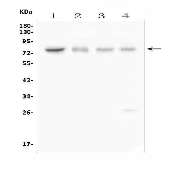
Figure 1. Western blot analysis of ABCG8 using anti-ABCG8 antibody (A01482-2).
Electrophoresis was performed on a 5-20% SDS-PAGE gel at 70V (Stacking gel) / 90V (Resolving gel) for 2-3 hours. The sample well of each lane was loaded with 50ug of sample under reducing conditions.
Lane 1: human A431 whole cell lysates
Lane 2: rat small intestine tissue lysates
Lane 3: mouse liver tissue lysates
Lane 4: rat RH35 whole cell lysates
After Electrophoresis, proteins were transferred to a Nitrocellulose membrane at 150mA for 50-90 minutes. Blocked the membrane with 5% Non-fat Milk/ TBS for 1.5 hour at RT. The membrane was incubated with rabbit anti-ABCG8 antigen affinity purified polyclonal antibody (Catalog # A01482-2) at 0.5 μg/mL overnight at 4°C, then washed with TBS-0.1%Tween 3 times with 5 minutes each and probed with a goat anti-rabbit IgG-HRP secondary antibody at a dilution of 1:10000 for 1.5 hour at RT. The signal is developed using an Enhanced Chemiluminescent detection (ECL) kit (Catalog # EK1002) with Tanon 5200 system. A specific band was detected for ABCG8 at approximately 76KD. The expected band size for ABCG8 is at 76KD.

Click image to see more details
Figure 2. Flow Cytometry analysis of HepG2 cells using anti-ABCG8 antibody (A01482-2).
Overlay histogram showing HepG2 cells stained with A01482-2 (Blue line). To facilitate intracellular staining, cells were fixed with 4% paraformaldehyde and permeabilized with permeabilization buffer. The cells were blocked with 10% normal goat serum. And then incubated with rabbit anti-ABCG8 Antibody (A01482-2,1μg/1x106 cells) for 30 min at 20°C. DyLight®488 conjugated goat anti-rabbit IgG (BA1127, 5-10μg/1x106 cells) was used as secondary antibody for 30 minutes at 20°C. Isotype control antibody (Green line) was rabbit IgG (1μg/1x106) used under the same conditions. Unlabelled sample without incubation with primary antibody and secondary antibody (Red line) was used as a blank control.

Click image to see more details
Figure 3. Flow Cytometry analysis of RH35 cells using anti-ABCG8 antibody (A01482-2).
Overlay histogram showing RH35 cells stained with A01482-2 (Blue line). To facilitate intracellular staining, cells were fixed with 4% paraformaldehyde and permeabilized with permeabilization buffer. The cells were blocked with 10% normal goat serum. And then incubated with rabbit anti-ABCG8 Antibody (A01482-2,1μg/1x106 cells) for 30 min at 20°C. DyLight?488 conjugated goat anti-rabbit IgG (BA1127, 5-10μg/1x106 cells) was used as secondary antibody for 30 minutes at 20°C. Isotype control antibody (Green line) was rabbit IgG (1μg/1x106) used under the same conditions. Unlabelled sample without incubation with primary antibody and secondary antibody (Red line) was used as a blank control.

Click image to see more details
Figure 4. IF analysis of ABCG8 using anti-ABCG8 antibody (A01482-2).
ABCG8 was detected in immunocytochemical section of HEPG2 cells. Enzyme antigen retrieval was performed using IHC enzyme antigen retrieval reagent (AR0022) for 15 mins. The cells were blocked with 10% goat serum. And then incubated with 5μg/mL rabbit anti-ABCG8 Antibody (A01482-2) overnight at 4°C. DyLight®488 Conjugated Goat Anti-Rabbit IgG (BA1127) was used as secondary antibody at 1:100 dilution and incubated for 30 minutes at 37°C. The section was counterstained with DAPI. Visualize using a fluorescence microscope and filter sets appropriate for the label used.
Protein Target Info & Infographic
Gene/Protein Information For ABCG8 (Source: Uniprot.org, NCBI)
Gene Name
ABCG8
Full Name
ATP-binding cassette sub-family G member 8
Weight
75679 MW
Superfamily
ABC transporter superfamily
Alternative Names
ATP-binding cassette sub-family G member 8; ATP-binding cassette, sub-family G (WHITE), member 8 (sterolin 2); ATP-binding cassette, sub-family G (WHITE), member 8; GBD4ATP-binding cassette, subfamily G, member 8; MGC142217; sterolin 2; sterolin-2; STSL ABCG8 GBD4, STSL, STSL1 ATP binding cassette subfamily G member 8 ATP-binding cassette sub-family G member 8|ATP-binding cassette, sub-family G (WHITE), member 8|sterolin 2
*If product is indicated to react with multiple species, protein info is based on the gene entry specified above in "Species".For more info on ABCG8, check out the ABCG8 Infographic

We have 30,000+ of these available, one for each gene! Check them out.
In this infographic, you will see the following information for ABCG8: database IDs, superfamily, protein function, synonyms, molecular weight, chromosomal locations, tissues of expression, subcellular locations, post-translational modifications, and related diseases, research areas & pathways. If you want to see more information included, or would like to contribute to it and be acknowledged, please contact [email protected].
Specific Publications For Anti-ABCG8 Antibody (A01482-2)
Hello CJ!
A01482-2 has been cited in 1 publications:
*The publications in this section are manually curated by our staff scientists. They may differ from Bioz's machine gathered results. Both are accurate. If you find a publication citing this product but is missing from this list, please let us know we will issue you a thank-you coupon.
Huang J,Wang Q,Chen M,Bi Y,Shi H,Zhou K. Effects of psoralen on hepatic bile acid transporters in rats. Hum Exp Toxicol.2020 Dec 15:960327120979346.doi:10.1177/0960327120979346. Epub ahead of print.PMID:33317360.
Species: Rat
A01482-2 usage in article: APP:WB, SAMPLE:LIVER TISSUE, DILUTION:1:500
Recommended Resources
Here are featured tools and databases that you might find useful.
- Boster's Pathways Library
- Protein Databases
- Bioscience Research Protocol Resources
- Data Processing & Analysis Software
- Photo Editing Software
- Scientific Literature Resources
- Research Paper Management Tools
- Molecular Biology Software
- Primer Design Tools
- Bioinformatics Tools
- Phylogenetic Tree Analysis
Customer Reviews
Have you used Anti-ABCG8 Antibody?
Submit a review and receive an Amazon gift card.
- $30 for a review with an image
0 Reviews For Anti-ABCG8 Antibody
Customer Q&As
Have a question?
Find answers in Q&As, reviews.
Can't find your answer?
Submit your question
1 Customer Q&As for Anti-ABCG8 Antibody
Question
We are currently using anti-ABCG8 antibody A01482-2 for human tissue, and we are well pleased with the ELISA results. The species of reactivity given in the datasheet says human, mouse, rat. Is it possible that the antibody can work on zebrafish tissues as well?
Verified Customer
Verified customer
Asked: 2019-08-13
Answer
The anti-ABCG8 antibody (A01482-2) has not been validated for cross reactivity specifically with zebrafish tissues, though there is a good chance of cross reactivity. We have an innovator award program that if you test this antibody and show it works in zebrafish you can get your next antibody for free. Please contact me if I can help you with anything.
Boster Scientific Support
Answered: 2019-08-13
