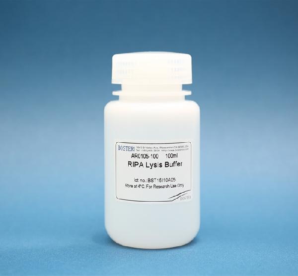
Product Info Summary
| SKU: | AR0105-100 |
|---|---|
| Size: | 100ml |
Product info
Overview
| Form Supplied | Ready-to-use 1X solution |
|---|---|
| Physical State | Liquid |
| Pack Size | 100 mL |
| Content | 50mM Tris•HCl pH 7.6, 150mM NaCl, 1% NP-40, 0.5% sodium deoxycholate, 0.1% SDS |
| Recommended working concentration | 10 mL RIPA Lysis Buffer per gram of tissue 0.5 mL RIPA Lysis Buffer per 5.0x10⁶ cells in suspension 0.5 mL RIPA Lysis Buffer per 5.0x10⁶ adherent mammalian cells |
| Storage & Expiration | Upon receipt store at 4°C. RIPA Lysis Buffer is stable for one year. Product is shipped on ice. |
| Assays per kit | 200 assays for 5.0x10⁶ cells 100 assays for 0.1g tissue |
| Compatibility with reagents | Fully compatible with Broad Spectrum Protease Inhibitor Cocktail and Broad Spectrum Phosphatase Inhibitor Cocktail |
| Equivalent | Thermofisher (Product No. 89900, 89901), Millipore Sigma (Product No. R0278) |
| Reagent Type | Western Blotting related reagent; Cell lysis buffer; Universal tissue extraction buffer; Detergent solution |
| Usage | Extraction of cytoplasmic, membrane and nuclear proteins |
| Cite This Product | RIPA Lysis Buffer (Boster Biological Technology, Pleasanton CA, USA, Catalog # AR0105-100) |
Description
RIPA Lysis Buffer is a complete cell lysis solution reagent used for rapid and efficient total cell lysis and solubilization of proteins from both adherent and suspension cultured mammalian cells, effectively extracting cytoplasmic, nuclear and membrane proteins. Protein lysis can be finished in 60 minutes.
Background
RIPA lysis extraction buffer contains non-ionic and ionic detergents which are able to extract protein from wide variety of cell types and membrane structures. RIPA buffer ensures efficient cell lysis and protein solubilization preventing protein degradation and interference with protein immunoreactivity and biological activity. Since most antibodies and protein antigens are not adversely affected by the components of this solution, RIPA buffer-conducted protein extraction is compatible with various downstream immunoprecipitation and molecular pull-down assays, including reporter assays, protein assays, immunoassays and protein purification. RIPA buffer reagent minimizes non-specific protein-binding interactions to keep background low, while allowing most specific interactions to occur, enabling studies of relevant protein-protein interactions.
Important Product Information
• If desired, add protease inhibitor (Product No. AR1182) and phosphatase inhibitor (Product No. AR1183) to the lysis buffer to prevent proteolysis and maintain phosphorylation status of proteins.
• Some protein kinases and other enzymes may be sensitive to the components of the RIPA Lysis Buffer, resulting in their decreased activity. In such cases, prepare a RIPA Lysis Buffer that does not contain sodium deoxycholate and SDS.
Additional Materials Required
• Protease inhibitor (Product No. AR1182) and phosphatase inhibitor (Product No. AR1183)
• 2 ml microcentrifuge tubes
• Tissue homogenizer
• Microcentrifuge capable of spinning at 10,000 x g
• Cell scraper
Procedure for Lysis of Monolayer-cultured Adherent Mammalian Cells
Note: Pre-chill an appropriate volume of RIPA Lysis Buffer at 4°C. If desired, add protease inhibitor and phosphatase inhibitor to the lysis buffer immediately before use.
1. In a microcentrifuge tube, resuspend 5×10⁶ cells in the growth media by scraping the cells off the surface of the plate with a cell scraper. Centrifuge harvested cell suspension at 600xg for 5min, then carefully remove and discard the supernatant.
2. Resuspend the cells in chilled PBS. Centrifuge at 600xg for 5min, then carefully remove and discard the supernatant.
3. Add 0.5 mL of chilled RIPA lysis buffer to the cell pellet. Vortex briefly. Incubate on ice for 30 minutes.
4. Centrifuge samples at 14000xg for 10 minutes.
5. Transfer supernatant to a new tube for further analysis.
Note: RIPA lysis buffer can be added directly to the flask containing cells. Please see the following procedures.
1. Carefully remove culture medium from adherent cells.
2. Wash cells with chilled PBS. Carefully remove PBS.
3. Add chilled RIPA lysis buffer to the cells. Vortex briefly. Incubate on ice for 30 minutes. (For the volume of the lysis buffer, follow the instructions listed below)
| SIZE of the plate/surface area | Volume of the lysis buffer |
| 100mm | 500-1000μL |
| 60mm | 250-500μL |
| 6-well plate | 200-400μL per well |
| 24-well plate | 100-200μL per well |
| 96-well plate | 50-100μL per well |
4. Centrifuge samples at 14000xg for 10 minutes.
5. Transfer supernatant to a new tube for further analysis.
Procedure for Lysis of Suspension-cultured Mammalian Cells
Note: Pre-chill an appropriate volume of RIPA Lysis Buffer at 4°C. If desired, add protease inhibitor and phosphatase inhibitor to the lysis buffer immediately before use.
1. In a microcentrifuge tube, harvest 5×10⁶ cells by centrifugation at 600xg for 5min. Carefully remove and discard the supernatant.
2. Resuspend the cells in chilled PBS. Centrifuge at 600xg for 5min, then carefully remove and discard the supernatant.
3. Add 0.5 mL of chilled RIPA lysis buffer to the cell pellet. Vortex briefly. Incubate on ice for 30 minutes.
4. Centrifuge samples at 14000xg for 10 minutes.
5. Transfer supernatant to a new tube for further analysis.
Procedure for Lysis of Tissues
Note: Pre-chill an appropriate volume of RIPA Lysis Buffer at 4°C. If desired, add protease inhibitor and phosphatase inhibitor to the lysis buffer immediately before use.
1. Place the fresh tissue into chilled PBS and rinse several times. Mince the tissue into small pieces.
2. Add RIPA Lysis Buffer to the tissue at 10:1. (i.e., Add 10 mL chilled lysis buffer per gram of tissue.) Use a smaller volume of reagent if a more concentrated protein extract is required.
3. Homogenize for several minutes at high speed until no tissue chunks remain.
4. Incubate on ice for 30 minutes.
5. Centrifuge at ~10000 x g for 10 minutes.
6. Transfer supernatant to a new tube for further analysis.
Precautions
• All steps of protein lysis should be operated on ice or at 4°C.
• Use BCA Protein Assay kit (Product No. AR0146) to quantify lysed proteins. Bradford Protein Assay kit is not recommended.
• There might be some transparent gel complex containing genomic DNA in lysed proteins. The protein fractions can be used for further analysis after centrifugation if target proteins have little connection with genomic DNA. When detecting target proteins related closely to genomic DNA, sonicate gel complex and then centrifuge to collect supernatant for further analysis. Common transcription factors such as NFKB, p53 can be detected without sonication.
Product Images
Validation Images & Assay Conditions

Click image to see more details
Specific Publications For AR0105-100
Hello CJ!
AR0105-100 has been cited in 76 publications:
*The publications in this section are manually curated by our staff scientists. They may differ from Bioz's machine gathered results. Both are accurate. If you find a publication citing this product but is missing from this list, please let us know we will issue you a thank-you coupon.
Menin mediates Tat-induced neuronal apoptosis in brain frontal cortex of SIV-infected macaques and in Tat-treated cells
Babao Dan inhibits the migration and invasion of gastric cancer cells by suppressing epithelial–mesenchymal transition through the TGF-β/Smad pathway:
Design, synthesis and structure-activity relationship optimization of phenanthridine derivatives as new anti-vitiligo compounds
Qu Y,Dou P,Hu M,Xu J,Xia W,Sun H.circRNA‑CER mediates malignant progression of breast cancer through targeting the miR‑136/MMP13 axis.Mol Med Rep.2019 Apr;19(4):3314-3320.doi:10.3892/mmr.2019.9965.Epub 2019 Feb 18.PMID:30816475.
Species: Human
AR0105-100 usage in article: APP:WB, SAMPLE: MCF-7 CELL AND ZR-75-30 CELL, DILUTION:NA
Lv J,Liu C,Chen FK,Feng ZP,Jia L,Liu PJ,Yang ZX,Hou F,Deng ZY.M2‑like tumour‑associated macrophage‑secreted IGF promotes thyroid cancer stemness and metastasis by activating the PI3K/AKT/mTOR pathway.Mol Med Rep.2021 Aug;24(2):604.doi:10.3892/mmr.2021.12249.Epub 2021 Jun 29. PMID:34184083.
Species: Human
AR0105-100 usage in article: APP:WB, SAMPLE:THP-1 CELL AND C643 CELL, DILUTION:NA
Chong Liu,Minmin Zhang,Shenyi Ye,Chenliang Hong,Jiaxi Chen,Ruyue Lu,Bingjie Hu,Weijun Yang,Bo Shen,Zhengyi Gu,"Acacetin Protects Myocardial Cells against Hypoxia-Reoxygenation Injury through Activation of Autophagy",Journal of Immunology Research,vol.2021,Article ID 9979843,12 pages,2021.https://doi.org/10.1155/2021/9979843
Species: Rat
AR0105-100 usage in article: APP:WB, SAMPLE:H9C2 CELL, DILUTION:NA
Chao Hu,Xiaobin Zhu,Taogen Zhang,Zhouming Deng,Yuanlong Xie,Feifei Yan,Lin Cai," Tanshinone IIA Inhibits Osteosarcoma Growth through a Src Kinase-Dependent Mechanism",Evidence-Based Complementary and Alternative Medicine,vol.2021,Article ID 5563691,15 pages,2021.https://doi. org/10.1155/2021/5563691
Species: Human,Mouse
AR0105-100 usage in article: APP:WB, SAMPLE:U2-OS CELL AND MG-63 CELL, DILUTION:NA
Xu J,Wang J,He Z,Chen P,Jiang X,Chen Y,Liu X,Jiang J.LncRNA CERS6-AS1 promotes proliferation and metastasis through the upregulation of YWHAG and activation of ERK signaling in pancreatic cancer.Cell Death Dis.2021 Jun 24;12(7):648.doi:10.1038/s41419-021-03921-3.PMID:34168120.
Species: Human
AR0105-100 usage in article: APP:WB, SAMPLE:PC TISSUE,ASPC-1 CELL, BXPC-3 CELL AND HPDE CELL, DILUTION:NA
Yang LG,Wang AL,Li L,Yang H,Jie X,Zhu ZF,Zhang XJ,Zhao HP,Chi RF,Li B,Qin FZ,Wang JP,Wang K.Sphingosine-1-phosphate induces myocyte autophagy after myocardial infarction through mTOR inhibition.Eur J Pharmacol.2021 Jun 15:174260.doi:10.1016/j.ejphar.2021.174260.Epub ahead of print.PMID:34144026.
Species: Rat
AR0105-100 usage in article: APP:WB, SAMPLE:LV TISSUE, DILUTION:NA
Huang X,Pei W,Ni B,Zhang R,You H.Chondroprotective and antiarthritic effects of Galangin in Osteoarthritis: An in Vitro and in Vivo Study.Eur J Pharmacol.2021 Jun 3:174232.doi:10.1016/j. ejphar.2021. 174232.Epub ahead of print.PMID:34090897.
Species: Rat
AR0105-100 usage in article: APP:WB, SAMPLE:CHONDROCYTES, DILUTION:NA
Customer Reviews
Have you used RIPA Lysis Buffer?
Submit a review and receive an Amazon gift card.
- $30 for a review with an image
0 Reviews For RIPA Lysis Buffer
Customer Q&As
Have a question?
Find answers in Q&As, reviews.
Can't find your answer?
Submit your question
3 Customer Q&As for RIPA Lysis Buffer
Question
In the AR0105-100 protocol it mentions "transferring the supernatant to a new tube for further analysis." What the supernantant can be analyzed for and what type of anaylsis would be useful?"
Verified customer
Asked: 2023-01-06
Answer
For the RIPA Lysis Buffer (AR0105-100), the supernatant can be analyzed for reporter assays, protein assays, immunoassays(WB, IP) and protein purifications.
Boster Scientific Support
Answered: 2023-01-06
Question
What buffer is recommended to be used to isolate protein from tumor tissue?
Verified customer
Asked: 2021-01-26
Answer
RIPA Lysis Buffer (AR0105-100) is recommended to be used to isolate protein from tumor tissue.
Boster Scientific Support
Answered: 2021-01-26
Question
What is the recommended method of sample preparation in regards to the Human TNFa kit for isolating brain tumor tissue and serum? Would AR0105-100 suffice?
Verified customer
Asked: 2019-04-30
Answer
The Ripa lysis buffer would suffice for preparing the brain tissue. RIPA Lysis Buffer (AR0105-100) Please see our sample preparation guide: https://www.bosterbio.com/protocol-and-troubleshooting/elisa-sample-preparation-guide
Boster Scientific Support
Answered: 2019-04-30


