This website uses cookies to ensure you get the best experience on our website.
- Table of Contents
The immune system can identify and eliminate cancer cells through both innate and adaptive mechanisms; however, such antitumor responses can be inhibited by the microenvironment through a process known as immunosuppression.
The immune system is capable of recognizing and eliminating cancer cells via both innate and adaptive mechanisms; however, such antitumor responses can be suppressed by the microenvironment, a process known as immunosuppression. Cancer immunotherapy tries to enhance the anticancer immune response by manipulating both immunosuppressive and immunostimulatory processes. It is critical, therefore, to comprehend tumor-infiltrating immune cells and their involvement in tumor growth and control. The environment of the tissue is also critical, as the interaction between malignant and immune cells is dynamic during carcinogenesis, mediated by a network of cytokines, chemokines, and growth factors.
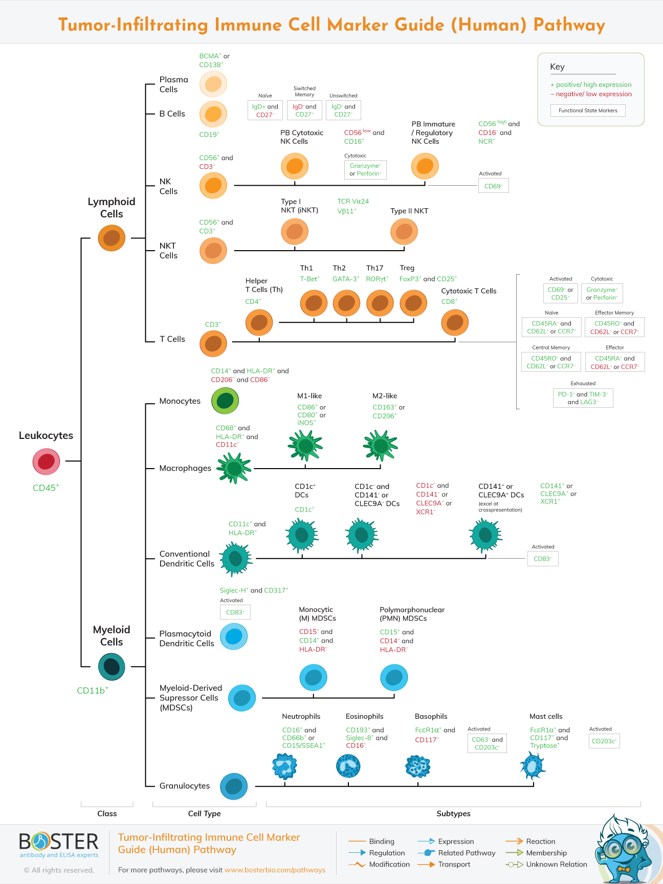
Reviewed below are the main immune system effector cells, their established cell-surface markers, and their function in cancer progression.
T cells are critical actors in the adaptive immune system; they are frequently identifiable by their CD3 expression and detect antigen via T cell receptors (TCRs), which recognize peptides presented by the major histocompatibility complex (MHC) (MHC). Circulating tumor cell antigens are transmitted to lymph nodes, where they are recognized by CD4+ and CD8+ T cells, also referred to as T helper and cytotoxic T cells. T helper cells release a range of cytokines following activation, including IFN. Cytotoxic T lymphocytes detect cells carrying tumor-specific antigens and kill them via apoptosis produced by perforin or granzyme.
T cell function is determined by the expression of a range of substances. Both CD69 and CD25 are increased in response to TCR signaling, but with differing kinetics, with CD69 increasing immediately after TCR ligation and CD25 increasing subsequently. T cell exhaustion, which is defined by impaired effector function and occurs during persistent infections and cancer, is defined by the expression of PD-1, TIM-3, and LAG3; however, these molecules are also elevated during T cell activation. Other T cell subtypes – such as naive, memory, and effector – are identified by a combination of CD45RA, CD45RO, and CD62L or CCR7. Multiple subtypes of CD4+ T lymphocytes secreting distinct cytokines — and eliciting distinct immunological responses – can be distinguished by their distinctive transcription factor expression. For instance, T-Bet, which is frequently expressed by Th1 cells, signals an anticancer phenotype and IFN production in general. FoxP3, which is produced by regulatory T cells (Treg), is associated with a protumor phenotype that suppresses the antitumor immune response via cytokine production and other mechanisms
Dendritic cells (DCs) are a component of the innate immune system that play a critical role in starting adaptive immunity by presenting antigens to activate naive T cells and secreting cytokines. DCs are classified into two subclasses: plasmacytoid and conventional. Plasmacytoid DCs are defined by the coexpression of Siglec-H and CD317 and are specialized in the production of significant levels of type I interferon, whereas conventional DCs are defined by the coexpression of CD11c and HLA-DR and are specialized in antigen presentation to T cells. Conventional DCs are further classified into those that express CD1c and enable CD4+ T cell activation and those that express CD141, XCR1, or CLEC9A and cross-present CD8+ T cells.
Macrophages are also innate immune cells; they are identifiable by their expression of CD68 and MHCII and their absence of CD11c. They are phagocytosis specialists and also produce cytokines that impact the immunological response. In general, macrophages are either pro-inflammatory (M1-like) or anti-inflammatory (M2-like). M1-like macrophages are characterized by the expression of CD80, CD86, or iNOS and boost antitumor immunity through phagocytosis of malignant cells and the generation of T cell activating ligands. M2-like macrophages, on the other hand, are characterized by the expression of CD163 or CD206 and can promote tumor growth by secreting immunosuppressive cytokines such as IL-10 and by encouraging a Th2 response. Additionally, M2 macrophages can express the immunosuppressive enzyme arginase, which depletes arginine in the tumor microenvironment, impairing T cell proliferation and function.
Finally, myeloid-derived suppressor cells (MDSCs) are a heterogeneous population of immature immunosuppressive cells found in a range of tumor types. Through the production of NOS2 and arginase 1, they have been demonstrated to suppress CD8+ T cell activation, stimulate Treg formation, and polarize macrophages toward an M2-like phenotype. MDSCs are classified into two main cell populations, monocytic and polymorphonuclear. Numerous unanswered concerns remain about these immunosuppressive cells, including their true differentiation from neutrophils and monocytes, the processes governing their accumulation and development, and their role in anticancer therapy resistance. MDSCs' particular markers are still being investigated. At the moment, they are most frequently identified by the presence of CD11b, the absence of HLA-DR, and the expression of either CD14 for monocytic MDSCs or CD15 for polymorphonuclear MDSCs..
Natural killer (NK) cells are the most common form of innate immune cell. They identify and eliminate cancer by recognizing the deregulation of MHC class I on tumor cells and/or the overexpression of ligands on tumor cells that bind to activating receptors on NK cells. NK cells are frequently identified by a combination of CD56 and CD16 expression and a lack of CD3.