This website uses cookies to ensure you get the best experience on our website.
- Table of Contents
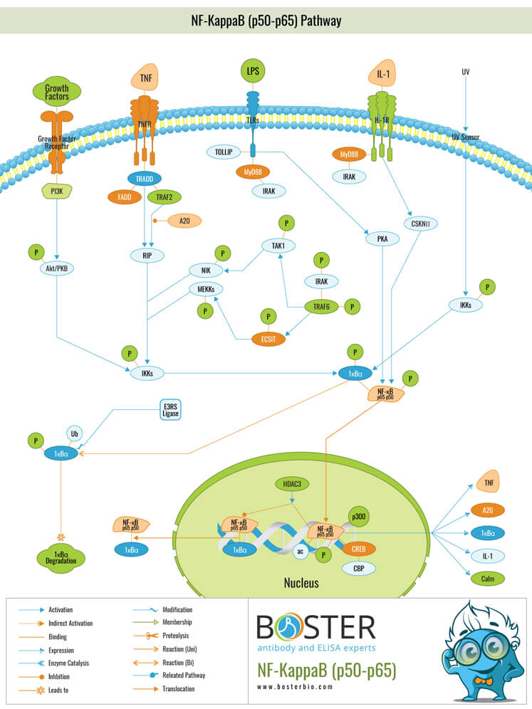
The NF-κB signaling system is a highly dynamic protein interaction network made up of components that regulate each other. The system includes five transcriptional monomers, two precursor proteins, three ankyrin repeat containing inhibitory IκB proteins, three stimulus-responsive inhibitory kinases (IKK complex: IKK-α and IKK-β, and IKK-γ or NF-κB essential modulator/NEMO), and ankyrin repeat containing regulatory proteins.( Basak S, Behar M, Hoffmann A. Lessons from mathematically modeling the NF-κB pathway. 2012)
The most abundant form of NF-κB activated by the canonical pathway is the heterodimer of p50 and p65. (Oeckinghaus A, Ghosh S. The NF-kappaB family of transcription factors and its regulation. 2009)
As stated, typically triggered by an NEMO-dependent activation of IKK, phosphorylation of IκB proteins followed by ubiquitination and degradation by proteasomes releases the NF-κB p65:p50 dimers from the inhibitory complex.( Collins P, Mitxitorena I, Carmody R. The ubiquitination of NF-κB subunits in the control of transcription. 2016)
The transcription factor nuclear factor kappa B (NF-B) is associated with inflammation. Without a stimulus, inhibitor of kappa B (IB) proteins suppress NF-B. When TNF, IL-1, and/or pathogen-associated molecular patterns (e.g. LPS) are stimulated, adaptor proteins such as MyD88 and TRAF signal for the activation of inhibitor of kappa B kinase (IBK), which then phosphorylates either IB (canonical pathway) or the p100 subunit of NF-B. (alternative pathway). The signal determines the activation pathway: TNF, IL-1, and TLR stimulation activate the classical pathway; CD40L and BAFF stimulation activate the alternative pathway.
The classical pathway involves ubiquitinating and then degrading phosphorylated IB, allowing NF-B to enter the nucleus and activate transcription of genes encoding cytokines such as TNF and IL-1. The alternative pathway involves activation of NF-B-inducing kinase (NIK), which activates IB kinase (IKK), resulting in phosphorylation of p100.
p65, alternatively referred to as RelA, is one of the five transcription factors that comprise the NF-B (nuclear factor kappa-light-chain-enhancer of activated B cells) family.The remaining members are RelB, c-Rel, and cleaved NF-B1 (p50) and NF-B2 proteins (p52). The most abundant heterodimer in the NF-B signaling pathway is formed by p50 and p65, which is found in the majority of cell types. In comparison, other members, such as c-Rel, are primarily expressed in hematopoietic cells (Napetschnig and Wu, 2013; Bassères and Baldwin, 2006).
p65, RelB, and c-Rel are defined by an N-terminal Rel homology domain that is combined with a C-terminal transactivating domain. NF-B1 and NF-B2 are, on the other hand, processed from large precursors (p105 and p100, respectively) into p50 and p52 subunits. This is a ubiquitin-mediated process that involves selective degrading ankyrin repeats from the C-terminal (Napetschnig and Wu, 2013; Bassères and Baldwin, 2006).
p65 is typically involved in the body's inflammatory response as a member of the NF-B signaling pathway. Stressful stimuli, such as free radicals, ultraviolet irradiation (UV), tumor necrosis factor (TNF), interleukin-1 beta (IL-1), pathogen-associated molecular patterns (PAMPs), or bacterial lipopolysaccharides, can activate this pathway (LPS). NF-B has been linked to memory and synaptic plasticity, while elevated levels of the protein have been linked to cancer (Courtois et al., 2006; Gutierrez and Davies, 2011).