Boster Pathways-> Gpcr, Calcium, Camp Signaling

Protein Kinase C Signaling Pathway

Protein Kinase C regulates a variety of cellular responses, including gene expression, protein secretion, cell proliferation and inflammation

Introduction to PKC

History of the Protein Kinase C

Protein kinase C (PKC) was first isolated from the brain by Yasutomi Nishizuka and his colleagues in 1977. It was obtained as a proenzyme that was switched on during proteolysis forming a serine and or threonine-protein kinase activity which is referred to as protein kinase M (PKM). The kinase activity of this enzyme has several functions. Enzyme interest was brought about by discovering that free calcium ions (Ca2+) and membrane phospholipids could together activate PKC even in the absence of proteolysis. This was done by Kishimoto in 1980.

Most Protein Kinase C function opinions are based on PKC activation assays based on the amount of membrane-bound Ca2+ and or phospholipids dependent kinase activity.

PKC Families

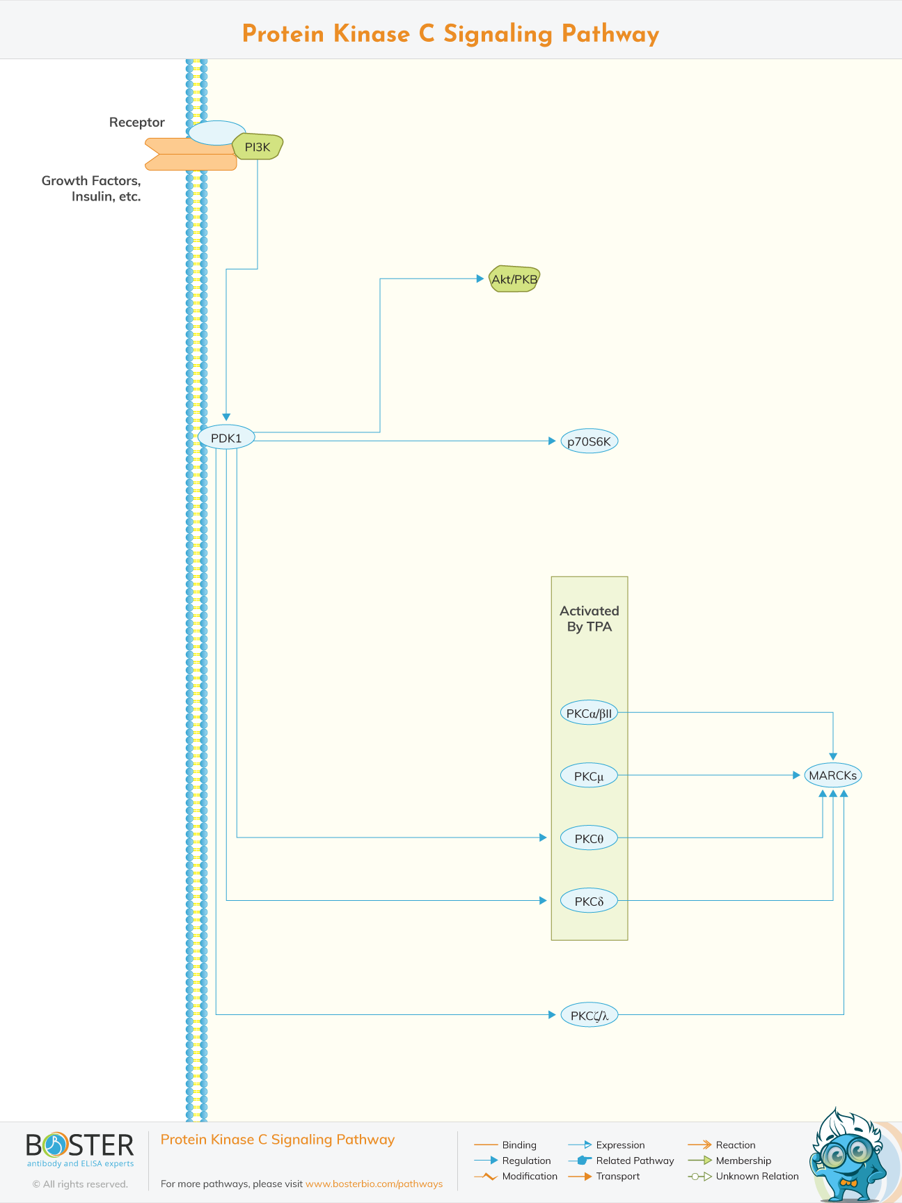
There are at least 10 members of the PKC family, which is classified into three subgroups: classical PKCs (alpha, betaI, betaII, gamma), new PKCs (delta, epsilon, eta, theta), and atypical PKCs (zeta, iota/lambda). The unique cofactor needs, tissue distribution, and cellular compartmentalization imply that each isoform has diverse roles and fine-tuned signaling cascades.

Mechanism Of Action

PKC is a family within cyclic AMP regulated kinases and cyclic GMP regulated kinases. They phosphorylate their substrate at specific sites which contain serine and threonine residues. PKCs are associated with two N-terminal domains C1 and C2 domains. C1 domain is sensitive towards phospholipids like diacylglycerols (DAGs). C2 domains are sensitive towards Ca2+. Some PKCs only have the C1 domains. But both types of PKCs are downstream effectors of the phospholipase C coupled receptors. PKC is also comprised of several isoforms like alpha, beta l and beta ll. Some of these isoforms regulate integrin affinity for extra cellular matrix (ECM) ligands. Integrins attach adhesion molecules like P- selectins upregulated during inflammation. Integrins are found on the surface of leukocytes.

Binding of integrins to the adhesion molecules like P- selectins allows for movement of leukocytes through the endothelium, a process called diapedesis. Outside in signaling of integrins can activate PKC isoforms leading to regulation of cell migration. For instance, a phospholipid product called p13k metabolites activates PKC regulating cell migration.

PKC that does not depend on Ca2+ or phospholipids for activation can also be found, it is this called a cognitive kinase.

After temporary activation dependent of say Ca2+ and independent PKC, some continue to remain active and are sovereign from Ca2+ and phospholipids. They stop cell migration by destroying receptors that contain serine and threonine residues.

PKC cause contraction of smooth muscles. They do so by phosphorylating an action binding protein called calponin. This reverses the inhibition of actin- activated myosin ATPase. This allows actin to interact with myosin thus increasing contraction of smooth muscles like vascular smooth muscles (VSM).

PKC Isoforms and their effects

  • PKCα (Protein Kinase C Alpha)

    PKC α is related to a variety of cell functions, including proliferation, apoptosis, differentiation, movement, and inflammation. However, the response induced by the activation or overexpression of PKCa varies depending on the cell type and sometimes the conditions. For example, in certain cell types, PKCa is involved in cell growth. Instead, it may play a role in cell cycle arrest and differentiation into other cell types. Therefore, the change in cellular response induced by PKC α is not an inherent characteristic of this isotype. The response is modulated by dynamic interactions with specific cell type factors: substrates, modulators, and anchor proteins.

  • PKCβ (Protein Kinase C Beta )

    Protein kinase Cβ (PKCβ) is a member of the lipid-activated serine / threonine PKC family and has been involved in a wide range of important cellular processes. Recently, the new role of PKCβ in the regulation of triglyceride homeostasis by regulating mitochondrial function has been explored. In this review, my purpose is to outline the relationship between PKCβ and lipid regulation and the recently gained knowledge about its role in energy homeostasis. Changes in the expression of fat PKCβ have been shown to be critical for diet-induced obesity and related metabolic abnormalities. A high-fat diet has been shown to induce PKCβ expression in target adipose tissue in an isotype and tissue specific manner.

  • PKCδ ( Protein Kinase C Delta)

    Protein kinase Cδ (PKCδ) exerts a variety of physiological functions through the ability to phosphorylate a variety of target proteins, which are involved in various cellular processes, such as signal transduction, apoptosis, proliferation and survival, transcription, hormonal regulation. and immune response. .

  • PKCε ( Protein Kinase C Epsilon)

    Protein kinase Cδ (PKCδ) exerts a variety of physiological functions through the ability to phosphorylate a variety of target proteins, which are involved in various cellular processes, such as signal transduction, apoptosis, proliferation and survival, transcription, hormonal regulation. and immune response.

    The importance of the role of PKCε has been described in the context of intracellular localization, because the selectivity and specificity of the substrate is achieved through the temporal and spatial targeting of PKCε. Therefore, PKCε regulates myocardial function under physiological and pathological conditions

  • PKCη (Protein Kinase C Eta)

    Protein kinase C (PKC) is a family of multiple genes that plays a key role in signal transduction and cell regulation. Ceta protein kinase (PKCη) is a unique member of the PKC family because its regulation is different from other PKC isoenzymes. PKCη has been shown to regulate cell proliferation, differentiation, and death. It has also been shown to contribute to the chemoresistance of various cancers. PKCη is associated with a variety of cancers, including renal cell carcinoma, glioblastoma, breast cancer, non-small cell lung cancer, and acute myeloid leukemia.

  • PKCθ (Protein kinase C theta)

    The protein kinase C theta (PKCθ) is a new Ca2 + independent member of the PKC subfamily that plays an important and non-redundant role in many aspects of T cell biology. Much progress has been made in understanding the role of PKCθ in the immune system and its unique translocation to immune synapses in stimulated T lymphocytes. Biochemical and genetic methods have shown that PKCθ is required for the activation and survival of mature T cells. Mutations in the PKCθ gene result in altered stimulation of AP1 receptor-induced transcription factors, NFκB, and NFAT, leading to defects in T cell activation and abnormal expression of apoptosis-related proteins, resulting in which results in a decrease in T cell survival.


FAQs

Protein kinase C (EC 2.7. 11.13) is a family of protein kinase enzymes that affect the activity of other proteins by phosphorylating the hydroxyl groups of serine and threonine amino acid residues on these proteins, or a member of this family.

PKC enzyme is activated in turn by signals, such as an increase in diglyceride (DAG) or calcium ion (Ca2 +) concentration. Therefore, PKC enzymes play an important role in several signal transduction cascades.

Protein kinases are an important class of intracellular enzymes that play a vital role in most signal transduction cascades, from controlling cell growth and proliferation to initiating and regulating immune responses.

CAMP binds to the R subunit, thereby inducing a conformational change, leading to the dissociation of the holoenzyme into a dimer of the R subunit and the free active C subunit. Any change in cAMP level will directly affect PKA function.

Protein Kinase (PTK) is an enzyme that regulates the biological activity of proteins by phosphorylating specific amino acids and using ATP as a source of phosphate, thereby inducing the conformational changes of proteins from inactive form to active form.