This website uses cookies to ensure you get the best experience on our website.
- Table of Contents
Ebola virus is an aggressive pathogen that infects humans and nonhuman primates, causing a highly lethal hemorrhagic fever syndrome. Typically, an Ebola virus infection lasts 14 to 21 days. At the start of the infection, nonspecific flu-like symptoms such as fever, myalgia, and malaise manifest. Patients develop severe bleeding and coagulation abnormalities as the infection progresses, including gastrointestinal bleeding, rash, and a variety of hematological abnormalities, such as lymphopenia and neutrophilia. When reticulo-endothelial cells come into contact with a virus, cytokines are released, which can result in exaggerated inflammatory responses that are not protective. When liver damage is combined with widespread viremia, disseminated intravascular coagulopathy results.
Ebola virus and the closely related Marburg virus are filoviruses, which are pleomorphic, negative-sense RNA viruses with the most similar genome organization to the Paramyxoviridae. The Ebola virus genome is 19 kilobytes in length and contains seven open reading frames that encode structural proteins such as the virion envelope glycoprotein (GP), nucleoprotein (NP), and matrix proteins VP24 and VP40; nonstructural proteins such as VP30 and VP35; and the viral polymerase. Through transcriptional editing, the Ebola virus GP open reading frame generates two gene products: a soluble 60–70 kDa dimeric soluble protein (sGP) and a full-length 150–170 kDa trimeric protein (GP) that inserts into the viral membrane . This envelope glycoprotein spike is expressed on the cell surface and is integrated into the virion to promote viral attachment and membrane fusion. Additionally, it has been demonstrated to be a critical factor in the pathogenicity of the Ebola virus . VP40 is found beneath the viral envelope, where it aids in the preservation of the virus's structural integrity. Due to its ability to induce its own release from cells in the absence of all other viral proteins, it has also been associated with late endosomes and is likely to mediate filovirus budding .
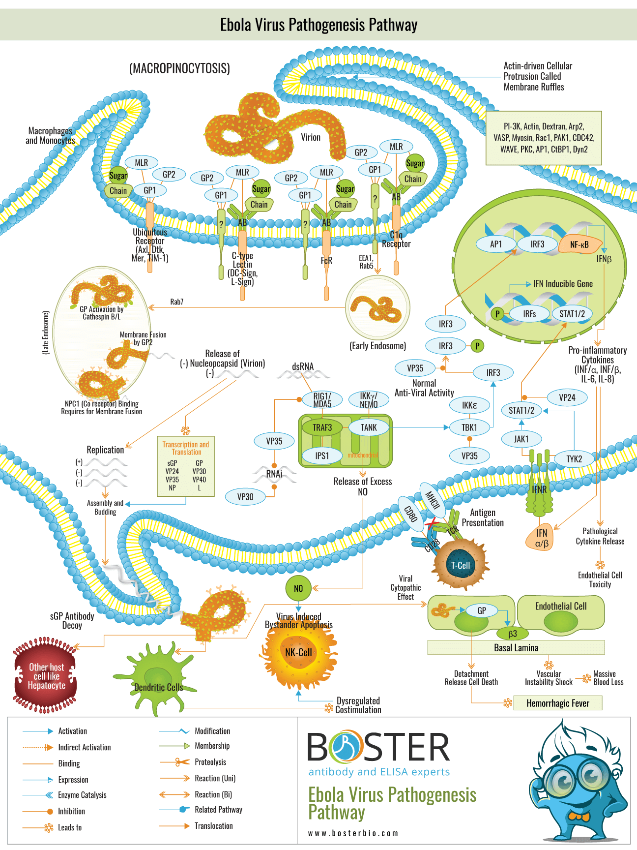
Entry into the host cell, as the first step in the viral life cycle, is a complex and multifaceted process. Although the mechanism of cellular entry is not fully understood, it involves uptake via a macropinocytosis-like mechanism. This mechanism is associated with actin polymerization-induced outward extensions of the plasma membrane. After fusion of the distal loop ends, these so-called membrane ruffles can fold back on themselves and form a macropinosome. The viral glycoprotein1 subunit (GP1) initiates virus entry into cells by interacting with both adherence factors and one or more receptors on the surface of host cells. The mucin-like region of GP is highly glycosylated with a high concentration of N- and O-linked glycans (MLR). The MLR plays a critical role in the entry of filoviruses into their preferred target cells . Numerous C-type lectins (e.g., DC-SIGN, L-SIGN) expressed on key cell types (e.g., macrophages, DCs) have been shown to mediate viral attachment and infection (Ref.8). TIM-1 acts as a receptor for virus entry on epithelial cells (Ref.6). Additionally, members of the Tyro3 receptor tyrosine kinase family (e.g., Axl, Dtk, and Mer) promote macropinocytic uptake induced by GP.
Internalized virus containing EBOV GP virions initially colocalizes with EEA1 (early endosomal antigen-1) positive compartments and is then trafficked to Rab5-positive early endosomes. At a later time point, colocalization of the virus with perinuclear Rab7/LAMP-1-positive late endosomes has been observed, and delivery to these compartments appears to be critical for entry (Ref.8). Within the acidified endosome, the highly glycosylated GP1 is cleaved to a smaller form by the low pH-dependent cellular proteases Cathepsin B and L, exposing RBS (residues in the receptor binding site), a ligand for NPC1 that mediates membrane fusion by the GP2 subunit. NCP1 is required for the fusion of the viral membrane and escape from the lysosomal vesicle . Apart from the conventional micropinocytosis mechanism of infection, Filoviruses utilize an alternative mechanism known as antibody-dependent enhancement (ADE) of viral infection, in which they enter cells in vitro via interaction between anti-GP antibodies and the cellular Fc receptor (FcR) or complement component C1q and its ligand, which likely promotes viral attachment to cells. The insertion of the EBOV GP2 fusion loop into host endosomal membranes is followed by the collapse of EBOV GP into a six-helix bundle, allowing for lipid mixing and hemifusion of host and viral membrane lipids. Finally, the hemifused host and viral membranes separate, forming a complete pore through which the viral genomic complex can enter the cytoplasm and continue the viral replication cycle .
Once the viral and internal cell membranes fuse, the virus particle uncoils and its anti-genome is transcribed into messenger RNA via nucleocapsid-associated viral proteins. The genome is transcribed by a complex consisting of VP30, VP35, and the viral polymerase L bound to an NP coated genome. Phosphorylation of VP30 causes it to dissociate from the VP35/L complex, signaling the transition from transcription to replication. NP, VP24, VP30, and VP35 replicate and coat virus genomes following this switch. L associates with the ribonucleoprotein complex during assembly via an interaction with VP35. The ribonucleoproteins then bind to the matrix protein VP40, and the viral particles are extruded through the plasma membrane via lipid raft microdomain regions. The expression and secretion of sGP acts as a decoy for antibodies against GP. The viral proteins VP35, VP30, and VP24 are expressed in all cell types and are involved in innate immune avoidance. VP35 inhibits RIG-I/MDA-5 signaling and interferon induction. Additionally, VP35 and VP30 inhibit the viral gene expression-specific RNAi response. VP24 inhibits Interferon (IFN) type I and II signaling. This inhibits Interferon-induced gene expression and, in antigen-presenting cells, prevents antigen presentation to T cells from being enhanced.