This website uses cookies to ensure you get the best experience on our website.
- Table of Contents
Insulin signals control cell growth and metabolic homeostasis. Dysregulation of this pathway can lead to metabolic diseases, such as diabetes.
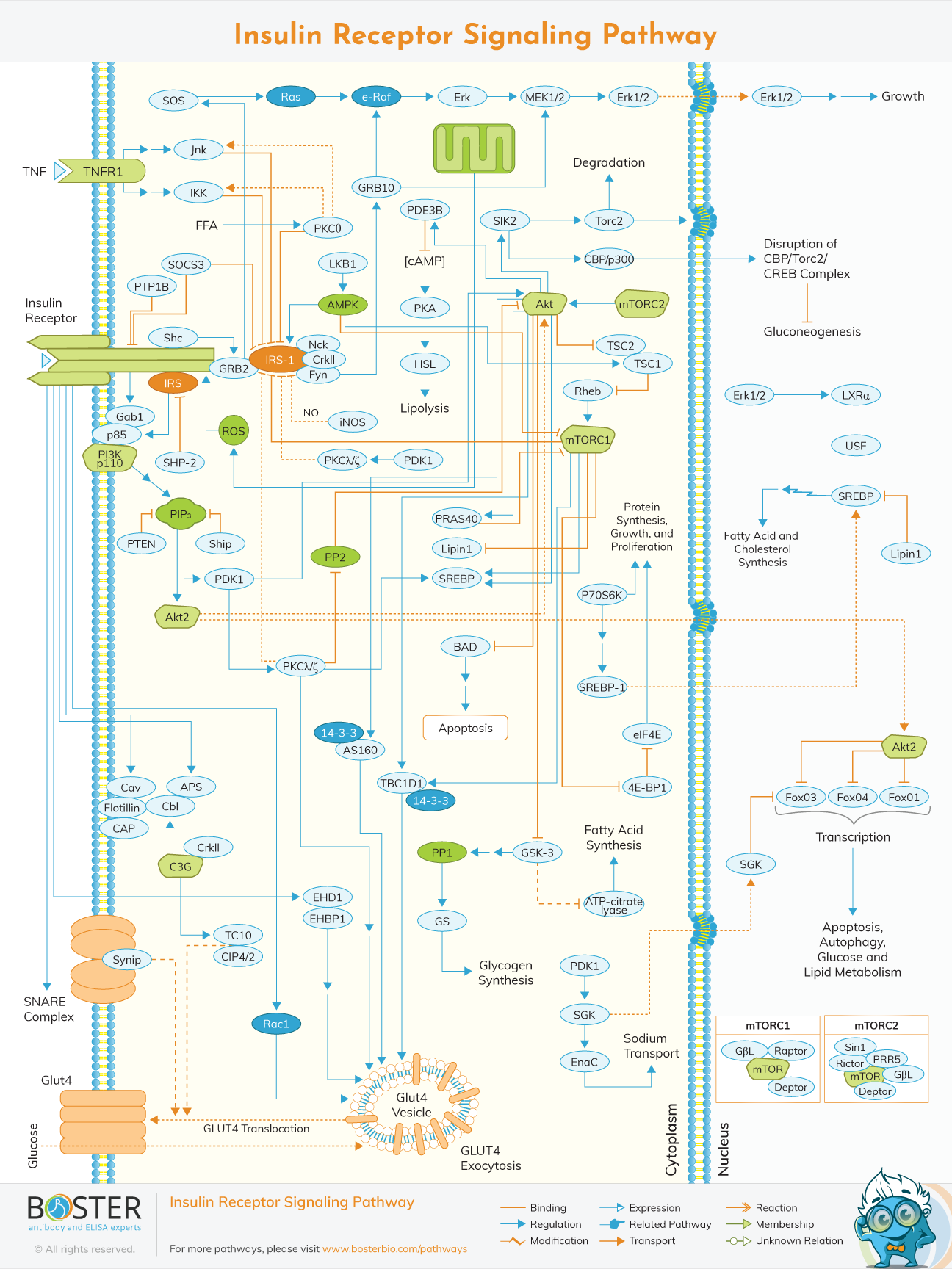
Insulin is a peptide hormone mainly used to lower blood sugar levels. It is secreted by beta cells in the pancreatic islets in response to nutrient absorption and elevated blood sugar levels. When insulin binds to receptors on target cells (such as skeletal muscle cells and adipocytes), the signal cascade begins, and finally the glucose transporter GLUT4 is transferred from the intracellular vesicles to the cell membrane. Once GLUT4 binds to the plasma membrane, it promotes the uptake of extracellular glucose, and then stores it as glycogen in these cells, thereby regulating blood sugar.
Insulin also regulates blood sugar by inhibiting gluconeogenesis (de novo production of glucose) and glycogenolysis (glycogenolysis) in the liver. In addition to regulating blood sugar levels, insulin also plays a key role in promoting protein and lipid synthesis and preventing the conversion of proteins and fats into glucose.Although insulin is considered a hormone that regulates glucose homeostasis, more and more studies clarify the broader role of this peptide. The insulin signaling pathway is usually very conservative.
The insulin receptor belongs to the superfamily of receptor tyrosine kinases (RTKs) and is activated by insulin and insulin-like growth factor (IGF12). It is a heterotetrameric protein consisting of two extracellular α subunits and two transmembrane β subunits, which are connected by disulfide bonds. Most RTKs bind directly to signal proteins. However, the insulin receptor binds to phosphorylated residues in associated proteins, a large family of docking proteins called the insulin receptor substrate family (IRS16), and the adapter protein Shc.
The insulin protein family combines several evolutionarily related active peptides: these include insulin, relaxin, insect pronuclides (bombyxin), insulin-like growth factors (IGF1 and IGF2), cell-specific insulin-like peptide mammalian Leydig ( INSL3 gene), early placental insulin-like peptide (ELIP), lobster insulin-related peptide (LIRP), mollusk insulin-related peptide (MIP) and Caenorhabditis elegans insulin-like peptide. The 3D structure of the different members of the family has been determined (Figure 3). The fold consists of two polypeptide chains (A and B) connected by two disulfide bonds: they all share the conservative arrangement of the four cysteines in the A chain, the first of which is connected to the third by a disulfide bond The second and the second and the fourth are connected to the cysteine in the B chain through interchain disulfide bonds.
The insulin receptor pathway plays a key role in regulating metabolic homeostasis. Insulin receptors act as tyrosine kinases and, by binding to insulin, initiate a phosphorylation cascade, increasing the concentration of glucose transport molecules in muscle and adipose tissue. By combining with the AKT / PI3K pathway to maintain greater regulation, this also promotes the conversion of glucose into glycogen for cellular storage.
When insulin binds to the extracellular α subunit of the insulin receptor, it will cause a conformational change, which then leads to autophosphorylation of various tyrosine residues present on the β subunit. They form the binding site of the IRS protein, which contains the phosphotyrosine binding domain (PTB), or the Shc adaptor protein, which contains the srchomology 2 (SH2) domain. The combination of the insulin receptor and IRS or Shc forms a platform that allows the assembly of signal transduction particles, thereby generating multiple intracellular signaling pathways.
Insulin Receptor-mediated signal transduction can be divided into IRS-mediated signal transduction pathway and non-IRS-mediated signal transduction pathway according to whether it mediates IRS (insulin receptor substrate).
Since the insulin signaling pathway is a complex pathway, many factors can regulate this pathway. Studies have found that protein tyrosine phosphatase 1B (PTP1B) plays a role in regulating the sensitivity of insulin signaling pathways and energy metabolism. Importantly, PTP1B knockout mice are more sensitive to the insulin signaling pathway and are resistant to obesity. Specific PTP1B inhibitors can significantly increase the body's sensitivity to insulin; αG (α glucose): inhibiting the activity of α-glucoside can slow down the production and absorption of glucose. Improve pancreatic sensitivity, protect pancreatic function, effectively prevent diabetes and improve complications. The emergence of aldose reductase (AR) is a restriction enzyme of glucose metabolism. It catalyzes the conversion of glucose to sorbitol, weakens the appearance of the insulin signaling pathway, and ultimately triggers diabetes; dipeptide kinase IV (DPPIV) can cleave and inactivate intestinal stimuli Insulin weakens the sensitivity of the insulin signaling pathway. In addition, the NFκB signaling pathway is closely related to the development of insulin resistance. Abnormal expression of NFκB and its related genes can directly or indirectly affect the transmission of insulin signaling pathway in different parts of the body through different pathways.