This website uses cookies to ensure you get the best experience on our website.
- Table of Contents
The hypoxia signaling pathway involves a hypoxic (low oxygen) environment that promotes cancer cell proliferation by preventing the degradation of Hypoxia Inducible Factor subunit α (HIF-α).
View pathway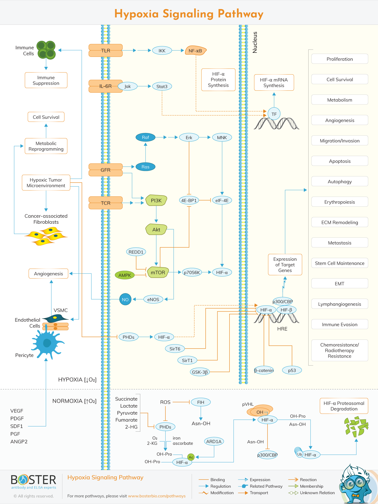
The hypoxia signaling pathway is mainly involved in the growth of cancer cells. A hypoxic (low oxygen) environment is a hallmark of cancer cell proliferation, which prevents the degradation of Hypoxia Inducible Factor subunit α (HIF-α).
In normal circumstances (normoxia), HIF-α is degraded by proteasomes, regulating its effects. However, in hypoxia, HIF-α degradation is inhibited, increasing its presence in the cellular environment. This allows HIF-α to dimerize with the β subunit (HIF-β), forming a transcriptional activator that promotes expression of HIF target genes.
In turn, this leads to several downstream effects that favor cancer cells, such as cell survival, proliferation, metastasis, immune cell evasion, resistance to chemotherapeutic drugs, angiogenesis, epithelial-mesenchymal transitions, and many others.
Guang L. Wang and Gregg L. Semenza were the first researchers to characterize and purify HIF-1 and identify its role in activating the erythropoietin gene (epo) in hypoxic conditions. This study was published in 1995 (Wang & Semenza, 1995).
Since then, HIF has been known as the master regulator of oxygen homeostasis, as it can activate many genes involved in hypoxia response. Other cell signaling pathways were also discovered that influence HIF activity, including the NF-κB, Jak-Stat3, and PI3K/Akt/mTOR pathways (Pouysségur, Dayan, & Mazure, 2006).
It is important to note that the hypoxia signaling pathway is only active in hypoxic circumstances (i.e. low oxygen concentration), which is typical in cancer cell microenvironments. In normal cases (normoxia), HIF-α activity is regulated through hydroxylation, ubiquitin tagging, and subsequent proteasome degradation.
However, during hypoxic conditions, PHDs and FIHs are less active. In turn, HIF-α accumulates in the cytoplasm, eventually translocating into the nucleus and binding to the constitutively-expressed HIF-β subunit as well as the transcriptional co-activator p300/CBP. The resulting protein complex binds to regulatory regions called hypoxia response elements (HREs), which leads to expression of downstream genes. In turn, this results in cell survival, proliferation, and angiogenesis, among other effects that confer tolerance to hypoxic conditions (Masson & Ratcliffe, 2014).
HIF activity can also be controlled indirectly, either through activation of HIF-α mRNA transcription or regulation of HIF-α protein synthesis. The former is achieved either through NF-κB signaling or the Jak-Stat3 pathway, while the latter occurs via PI3K/Akt/mTOR pathway. Either of these processes results in upregulation of HIF-α, promoting cell survival in hypoxic conditions (Brahimi-Horn, Chiche, & Pouysségur, 2007)
In normoxic conditions, HIF-α is regulated by prolyl hydroxylases (PHDs) and factors inhibiting HIF (FIHs). PHDs and FIHs are oxygen-dependent, so in normoxia, they are active and add hydroxyl groups to two specific proline residues in HIF-α. Similarly, FIH hydroxylates HIF-α at an asparagine site. The hydroxylated proline residues allow for the binding of the von Hippel-Lindau protein (pVHL), a ubiquitin ligase complex. Once HIF-α has been ubiquitinylated, it is subsequently degraded by a proteasome. This is how HIF activity is kept to a minimum during normal conditions (Gorres & Raines, 2010).
| Protein Target | Discussion |
|---|---|
| HIF | Hypoxia inducible factors (HIFs) are the main drivers of the hypoxia signaling pathway. It comprises two subunits - alpha and beta. There are three known alpha (1ɑ, 2ɑ, 3ɑ) subunits and one known beta (1β) subunit. When the alpha and beta subunits combine, they associate with a co-activator known as p300/CBP. The resulting complex binds to hypoxia response elements (HREs), activating the genes downstream that promote cell survival in hypoxic conditions. |
| FIH | Factor inhibiting HIF (FIH) is a 40-45 kDa protein that hydroxylates a specific asparagine site in HIF-α. Together with PHD, they cause HIF-α to be degraded through the ubiquitin-proteasome pathway. |
| PHD | Prolyl hydroxylase domain enzymes (PHDs) are tetramers with two subunits: a 59-kDa alpha subunit and a 55-kDa beta subunit. PHDs catalyze the hydroxylation of two specific proline residues in HIF-α, allowing the association of the von Hippel-Lindau tumor suppressor (pVHL). In turn, this causes ubiquitination of HIF-α and subsequent proteasome degradation. |
| pVHL | The von Hippel-Lindau tumor suppressor (pVHL) has two forms: one is 18 kDa in size and the other is 30 kDa. The protein exhibits E3 ubiquitin ligase activity, and it ubiquitinates HIF-α for degradation by proteasomes. |
The hypoxia signaling pathway is mostly involved in solid-tumor cancers. As tumor cells multiply, the oxygen supply of the surrounding environment decreases, leading to hypoxia. This condition activates the HIF-mediated hypoxia signaling pathway, which leads to the expression of several genes that cancer cells use to their advantage. These target genes lead to effects favorable to cancer cells, such as cell survival, proliferation, metastasis, epithelial-mesenchymal transition, immune suppression, and resistance to radiotherapy and chemotherapy. For these reasons, HIF and other molecules involved in the hypoxia signaling pathway are potential therapeutic targets (Lee, Ko, Ju, & Eltzschig, 2019).
In ischemic heart disease, cardiac muscles are deprived of an adequate supply of oxygen, leading to a loss of function and cell death in the affected areas. Due to the low oxygen levels, the hypoxia signaling pathway is activated, leading to increased levels of HIF in cardiac muscle cells. Several studies have shown that HIF upregulation during ischemic attacks provides a certain level of protection to the heart. Expression of the gene targets of HIF promotes survival of heart muscle cells during ischemic episodes (Lee, Ko, Ju, & Eltzschig, 2019).
A certain cancer drug known as Salidroside, when combined with platinum drugs such as cisplatin and oxaliplatin, was shown to promote the degradation of HIF-1α through the ubiquitin-proteasome pathway in both in vitro and in vivo models of hepatocellular carcinoma (Qin et al., 2018). Platinum drugs are commonly used for chemotherapy in certain cancers, but treatment is often hindered by drug resistance, which is conferred by genes expressed as a consequence of highly active HIF in hypoxic tumor microenvironments.
However, administering Salidroside along with platinum-based chemotherapeutic drugs increases the effectiveness of treatment. With the downregulation of HIF-1α, cancerous cells lose their resistance to the platinum drugs as well as the ability to undergo epithelial-mesenchymal transition. The latter, in effect, prevents metastasis of the cancer cells.
This study was limited to cell culture and mouse models. Thus, although a combination therapy of Salidroside plus platinum drugs is promising, it is yet to be tested on human patients.