This website uses cookies to ensure you get the best experience on our website.
- Table of Contents
CMV is used to determine the roles of regulatory elements and the effects of the mitogen-activated protein kinase (MAPK) pathways.
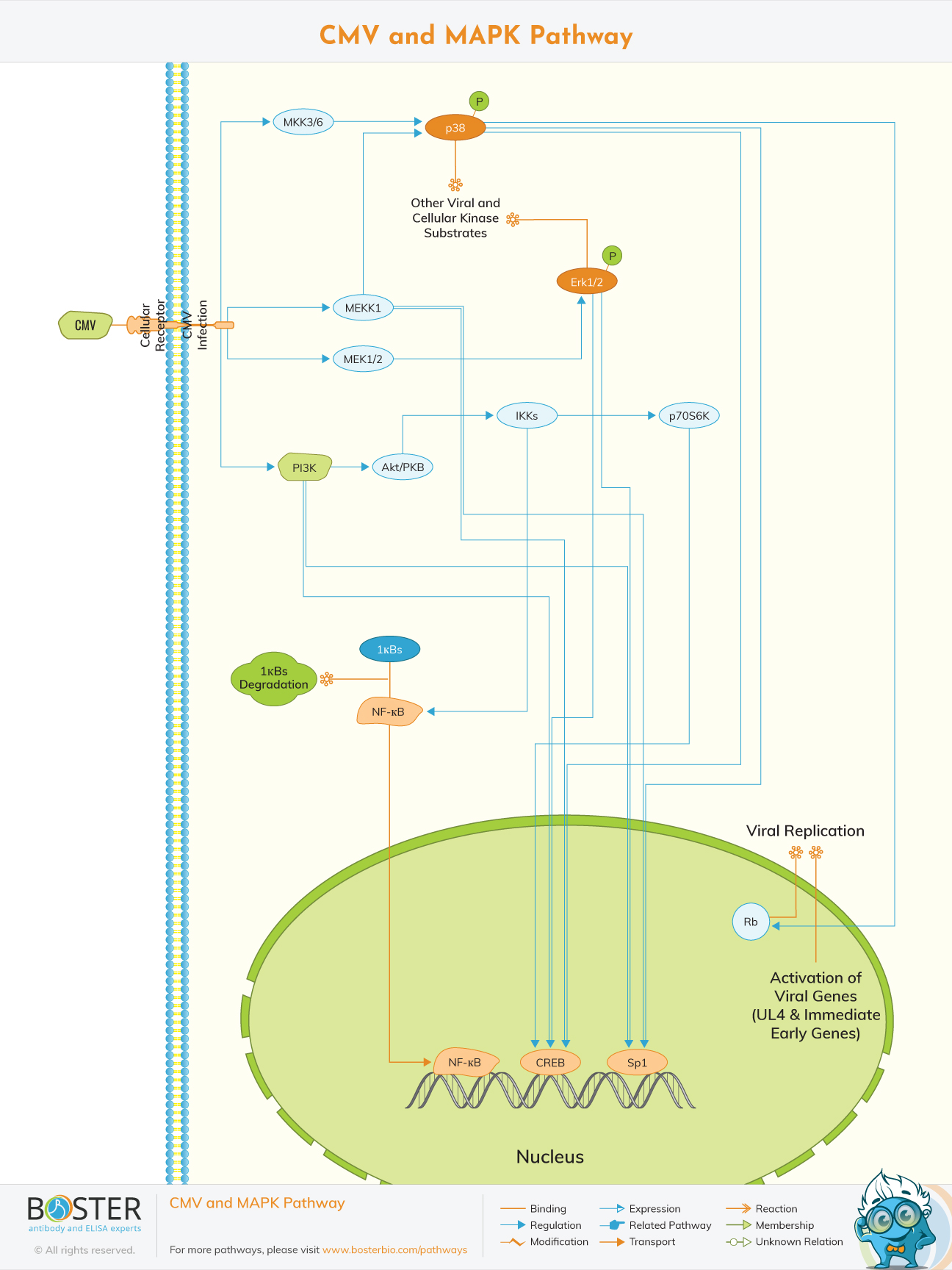
Cells respond to CMV (Cytomegalovirus) by invoking a cascade of responses resulting in signal transduction and regulation of cellular gene expression. CMV is a herpes virus that infects most cell types and establishes latency in leukocytes. A CMV infection is normally subclinical but can be fatal for persons with a weak immune system. CMV binds to cellular receptors on the cell membrane and enters a cell. Once this happens, a cell is said to be having a CMV infection. One of the early cellular responses to CMV infection is production of IP3 (Inositol 1,4,5-triphosphate) and DAG (1,2-Diacylglycerol) from PI3K (Phosphatidylinositol 3-Kinases) by phosphorylation. These lipids serve as second messengers and are able to regulate phosphorylation of a number of kinases, including Akt1/PKB (Protein Kinase-B) and p70S6K and p85S6K (Ribosomal S6 Kinases), respectively.
In top of that, the CMV particle in the host cell carry phosphatase activity and activate the MAPK (Mitogen-Activated Protein Kinase) pathway. MAPK activation by CMV leads to activation of transcription of viral genes, increasing the production of viral gene products.A CMV infection is usually asymptomatic but can be deadly in the immune system
The initial stage in CMV infection is attachment to a cellular target; however, CMV receptors have yet to be identified. It has been proposed that heparin sulfate and CD13 are involved in CMV attachment. The synthesis of IP3 (Inositol 1,4,5-triphosphate) and DAG (1,2-Diacylglycerol) from PI3K (Phosphatidylinositol 3-Kinases) by phosphorylation at the D-3 site is one of the early cellular responses to HCMV (Human CMV) infection. Once phosphorylated at the D-3 location, these lipids act as second messengers, regulating the phosphorylation of a variety of kinases, including Akt1/PKB (Protein Kinase-B), cAMP (cyclic Adenosine 3', 5'-Monophosphate)-dependent PKA (Protein Kinase-A), certain PKC (Protein Kinase-C), p70S6K and p85S6K (Ribosomal S6 Kinases), respectively.
Furthermore, the HCMV particle in the host cell has phosphatase activity, which activates the membrane-proximal PLC (Phospholipases-C) and A2 pathways, as well as the archetypal MAPK (Mitogen-Activated Protein Kinase) pathway. The MAPK signal transduction cascade is made up of three components that are conserved from yeast to humans.
CMV activation of MAPK causes transcription of viral genes to be activated, boosting the generation of viral gene products. MEKK1 controls the immediate early promoter indirectly via downstream kinase signaling and directly via NF-kappaB activation (Nuclear Factor-kappaB). The process of NF-kappaB activation includes the release of the transcription factor from the inhibitory subunit I-kappaB-Alpha (Inhibitor of Kappa Light Chain Gene Enhancer in B-Cells-Alpha) and is mediated by a GB-dependent mechanism via IKKs (I-KappaB Kinases). Later in the infection, different mechanisms amplify the early activation of NF-kappaB. This second wave of NF-kappaB activation is dependent on the activation of the CMV major immediate-early promoter by NF-kappaB. CMV-activated MAPK pathways result in enhanced transcription of viral genes and replication of the viral genome. Following infection, ERK1/2 (Extracellular Signal-Regulated Kinases) and p38 are activated, which controls viral gene activity via cellular transcription factors operating through the basal transcription elements and the viral UL4 promoter situated upstream. Rb (Retinoblastoma) is another target of sustained p38 activation during infection, which contributes to viral multiplication .
The E2F family of transcription factors is Rb's traditional binding partner, and hyperphosphorylation of pRb is required to relieve pRb-mediated repression of E2F, resulting in cell cycle advancement through the G1/S transition point. The HCMV IE2-86 protein, on the other hand, binds to pRb and alleviates pRb-mediated inhibition of E2F transactivation activity.