This website uses cookies to ensure you get the best experience on our website.
- Table of Contents
Apoptosis or programmed cell death is a mechanism which involves the mammalian system killing its own cells. This is of importance since it helps with the selective removal of aging, damaged, or otherwise unwanted cells from the body. Also, it’s important in various physiological processes such as embryogenesis, normal tissue development, and the immune response. Due to this, deregulation of apoptosis leads to pathological conditions such as carcinogenesis.
Apoptosis is involved in various diseases including myocardial ischemia, neurodegenerative diseases, stroke, septic shock, and Cancer.
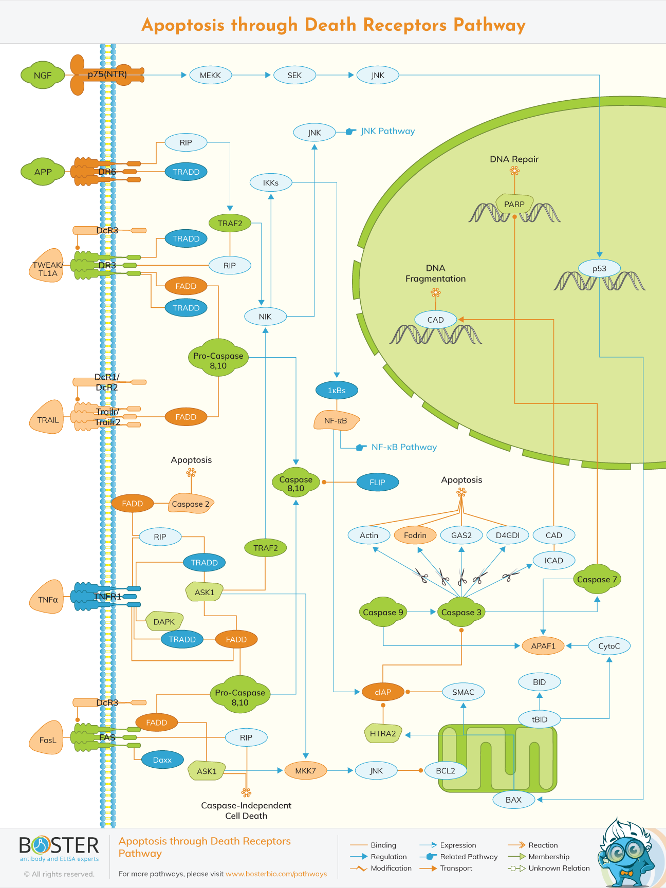
For the initiation of apoptosis, two major pathways are involved and these include; extrinsic death receptor pathways and intrinsic mitochondrial pathways. Apoptosis is generally mediated through the activation of caspases which are specific proteases that effectors of cell suicide. Caspases lead to disintegration of the cell structure thus forming apoptotic blebs and DNA fragmentation. These apoptotic bodies are then engulfed by phagocytic cells such as macrophages
Apoptosis originates from inside the cell in the mitochondria. This is activated by various stress stimuli such as DNA damage, heat, viral virulence factors, actions of some oncoproteins and tumor suppressor genes.
Cytosolic molecules sense these stress stimuli and then transduce the signals to the mitochondria which in turn leads to the alteration of the outer membrane of the mitochondria. In the case of DNA damage, ATM and CHK proteins sense this and thus leads to activation of P53. P53 recruits Bax and Bak proteins which cleaves the outer membrane of the mitochondria by forming pores. The loss of the integrity of the membrane leads to the release of mitochondrial proteins such as cytochrome c. Cytochrome c promotes the caspase cascade of cell execution and is known as apoptogenic factors. Other apoptogenic factors include; Omi/Htr2A and Smac/Diablo (which together with cytochrome c are released first after the disintegration of the outer membrane), and AIF and endoG (released after the subsequent damage of the inner membrane of the mitochondria).
Cytochrome c leads to the activation of Apoptosis Protease Activating Factor-1 (Apaf-1) and subsequent formation of apoptosome which is referred to as the ultimate cellular death machine. Apoptosome facilitates the activation of pro-caspase-9 to caspase-9 which is an auto-catalytic process. Despite this, apoptosome requires additional regulatory factors such as Smac/Diablo, in order to fully activate the caspase cascade. Smac/Diablo is a protein that interacts with IAPs (Inhibitor of Apoptosis Proteins), that inhibits activation of pro-caspase-9, and disintegrates them from their inhibitory function. Active caspase 9 activates pro-caspase-3 to caspase 3.
Caspase 3 activates protease and nuclease enzymes. Proteases break down proteins and protein structures in the cell and nuclease enzymes break down nucleic acids such DNA and RNA found in the nucleus hence leading to the disintegration of the cell. This disintegration leads to the formation of apoptotic blebs which are then engulfed by phagocytic cells such as macrophages.
Extrinsic pathway originates from outside the cell. This apoptotic pathway is activated by the binding of cytokine ligands to the Fas receptors which are also known as death receptors. These cytokine ligands include FasL, TNF, and TRAIL. The death receptors contain a specific intracellular domain bound to it known as Death Domain (DD).
Once cytokine ligand, FasL, binds to the Fas receptor this leads to the recruitment of an adaptor molecule known as Fas Associated Death Domain (FADD). FADD also contains a Death Effector Domain (DED) which is also an interaction Domain. DED of FADD interacts with the DED of pro-caspase-9 in order to form a protein complex that is known as Death Inducing Signaling Complex (DISC). DISC activates pro-caspase-8 to caspase 8. Pro-caspase-8 is the inactive form of caspase 8.
The caspase 8 leads to the activation of pro-caspase-3 to caspase 3. Caspase 3, a protein, is the effector caspase that leads to cell execution.
After it’s activation, caspase 3 degrades the inhibitors that are bound to the nuclease enzyme hence activating nuclease enzyme, Nuclease enzyme breaks down nucleic acids such as DNA and RNA found in the chromosomes.
On the other hand, caspase 8 also activates Bid protein (a member of the Bcl-2 family which is an anti-apoptotic member that inhibits Bax activity on the outer membrane of the mitochondria) to tBid which is a pro-apoptotic protein (induces apoptosis by promoting Bax activation).
This then leads to the triggering of the intrinsic mitochondrial pathway.