This website uses cookies to ensure you get the best experience on our website.
- Table of Contents
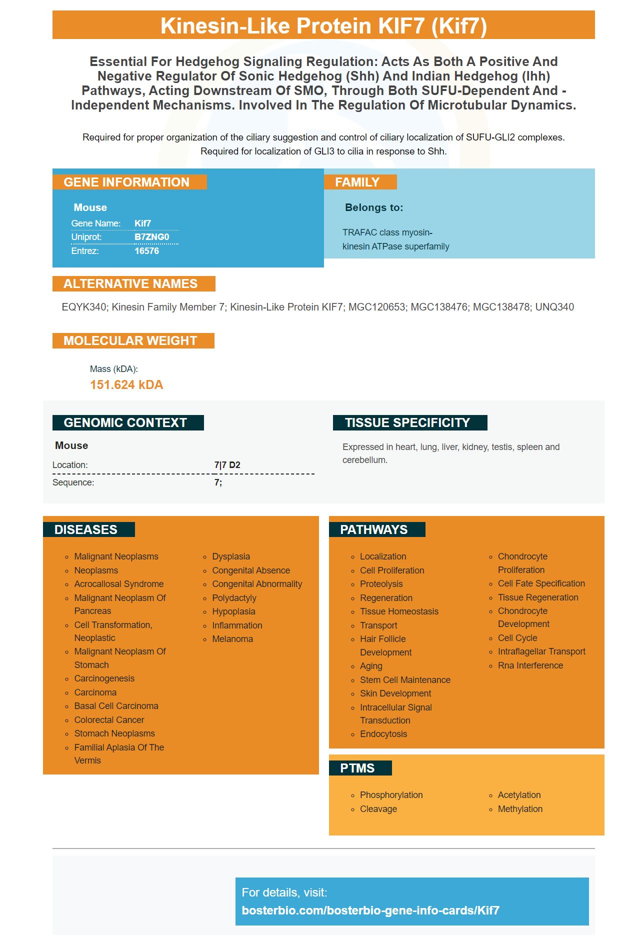
Facts about Kinesin-like protein KIF7.
Required for proper organization of the ciliary suggestion and control of ciliary localization of SUFU-GLI2 complexes. Required for localization of GLI3 to cilia in response to Shh.
Mouse | |
---|---|
Gene Name: | Kif7 |
Uniprot: | B7ZNG0 |
Entrez: | 16576 |
Belongs to: |
---|
TRAFAC class myosin-kinesin ATPase superfamily |
EQYK340; kinesin family member 7; kinesin-like protein KIF7; MGC120653; MGC138476; MGC138478; UNQ340
Mass (kDA):
151.624 kDA
Mouse | |
---|---|
Location: | 7|7 D2 |
Sequence: | 7; |
Expressed in heart, lung, liver, kidney, testis, spleen and cerebellum.
If you're interested in using KIF7 in your studies, then you've come to the right place. This article will discuss its use in biochemistry and pharmacy. It also discusses the importance and use of membrane staining in protein transfer, Autoradiography films, and DAB-chromogenic detection methods. Boster Bio, Best Uses and the KIF7Marker summarizes the most recent research regarding this protein.
A pooled sample containing 10mg of human serum was separated by SDS–PAGE and transferred onto PVDF membranes or nitrocellulose membranes. These membranes were then cut into five equal parts and fixed with acetone or 50% methanol. Subsequently, the samples were stained with LCA and SNA lectins. These samples were heated up to 100°C for 30 seconds, and then analyzed quantitatively.
The proteins were transferred from NC to PVDF membranes with an 8% SDS–PAGE gradient. The bound proteins were stained using direct blue-71. The band intensities of the bound proteins were quantified with GraphPad Prism version 6.
The sample comb should then be gently pressed onto the membrane to detect proteins. After pressing the sample comb carefully over the membrane, it should be incubated at a temperature of 37°C for between 30-60 minutes. It may be necessary to optimize the time of incubation. To remove any unbound antibodies, the membrane should first be washed three more times with TBS Wash Buffer. A secondary antibody should be added in a ratio of 3:1 to the membrane after it has been washed.
To quantify the transfer efficiency of proteins, the blots were incubated with antibodies, lectins, or proteins. To visualize the proteins bands, the Anti-mouse IgG MAb was used. Membranes were then set in a Canon scanner. The resulting images were then analyzed using densitometry. Images were then analyzed using Image J and GraphPad Prism version 6.
This marker has many applications. If you're not familiar with its uses, it is easy for you to find one. We will be discussing its use in biomedical science. This protein is a receptor of kinesin which is a nucleoprotein that controls the expression and function of several other genes. The marker is used to identify these genes, and it is derived from bovine chorionic gonadotropin (hs-Ca2+).
The ECL chemical chemiluminescent detection is a widely-used method for quantitatively copying proteins. It has some limitations. It is not possible for both enzymatic processes to be captured simultaneously. Multiplex PCR cannot be performed simultaneously or in every run. It is also impossible to quantify the amount of protein on a membrane. Researchers must therefore learn more about ECL limitations and how they can be overcome.
The binding of a primary antibody and a target protein is the first step in ECL chemistry. The secondary antibody is usually labeled with an enzyme (alkaline phosphatase and horseradish oxidase), which releases photons if it reacts with the target proteins. You can measure the photons produced by the reaction using either an xray film or digital imaging.
Enhanced chemiluminescence, a luminol based substrate, can be used for Western Blot applications. The amount of antigen in the substrate is proportional to the reaction between HRP-labeled antibody and the substrate produces a luminous signal. This indirect method of protein detection is further enhanced by the use of pictograms.
The first step is to prepare a high salt wash buffer containing 10 mM Tris (pH 7.4), 0.5 M NaCl and 1 mM NaH2PO4 with 0.1% SDS/Tween-20. The next step is to blot the membrane with the protein side facing up. The blot membrane should be placed on the film and exposed for one minute. Light emission is at its peak within five to thirty minutes of incubation of the substrate. Finally, a CCD detector is used to capture chemiluminescent images.
Boster Bio's DAB system for chromogenic detection is a powerful system. It integrates a unique technology called Intensification that uses phosphors to enhance chromogenic signals. This system uses horseradish peroxidase to catalyze deposition of two separate Linkers. These Linkers react with streptavidin-peroxidase conjugates, and then detect proteins using diaminobenzidine (DAB).
PMID: 19592253 by Endoh-Yamagami S., et al. The mammalian Cos2 homolog Kif7 plays an essential role in modulating Hh signal transduction during development.
PMID: 19666503 by Liem K.F. Jr., et al. Mouse Kif7/Costal2 is a cilia-associated protein that regulates Sonic hedgehog signaling.