This website uses cookies to ensure you get the best experience on our website.
- Table of Contents
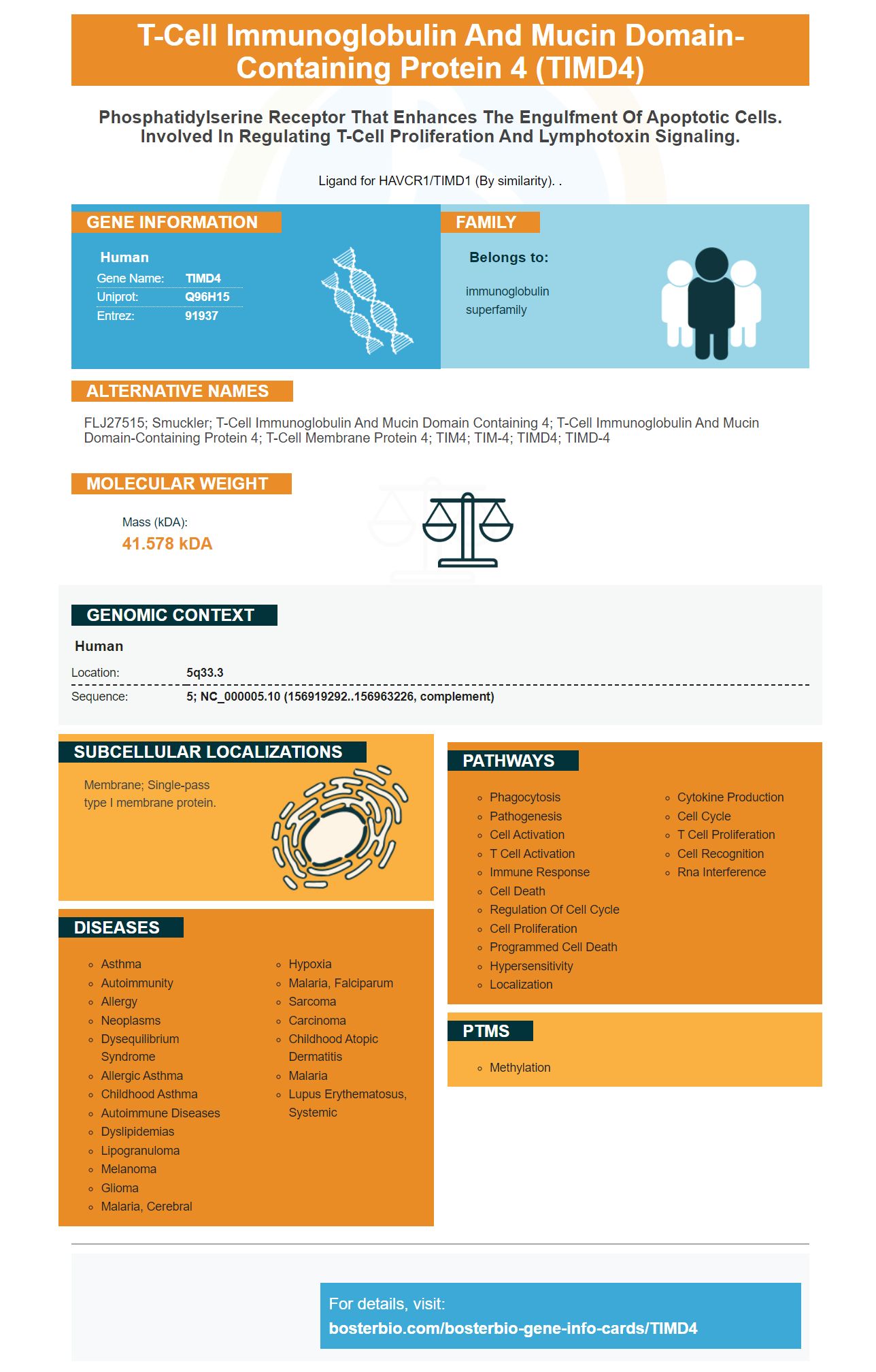
Facts about T-cell immunoglobulin and mucin domain-containing protein 4.
Ligand for HAVCR1/TIMD1 (By similarity). .
Human | |
---|---|
Gene Name: | TIMD4 |
Uniprot: | Q96H15 |
Entrez: | 91937 |
Belongs to: |
---|
immunoglobulin superfamily |
FLJ27515; Smuckler; T-cell immunoglobulin and mucin domain containing 4; T-cell immunoglobulin and mucin domain-containing protein 4; T-cell membrane protein 4; TIM4; TIM-4; TIMD4; TIMD-4
Mass (kDA):
41.578 kDA
Human | |
---|---|
Location: | 5q33.3 |
Sequence: | 5; NC_000005.10 (156919292..156963226, complement) |
Membrane; Single-pass type I membrane protein.
Boster Bio has primary antibodies against TIMD4 that are available. Developed in mice and rabbits, these antibodies can recognize TIMD4 in many animal samples. TIMD4 can recognize phosphoidylserine receptors that regulate lymphotoxin signaling. It also regulates T-cell proliferation. It also acts as a ligand to HAVCR1/TIMD1. In 2006, Ota and Schmutz sequenced 21,243 copies of the human cDNA and chromosome 5.
Biological assays that detect the protein use antibodies against TIMD4. Biological assays could use monoclonal (or polyclonal) antibodies that bind TIMD4 with animal samples. Boster Bio develops its antibodies to TIMD4 in mice and rabbits. TIMD4 acts as a phosphoidylserine and regulates T-cell growth and lymphotoxin response. It binds with the HAVCR1/TIMD1 signaling receptor and regulates Lymphotoxin signaling. Numerous scientific studies have found that the human gene coding TIMD4 is composed of 21243 codons. Accordingly, boster bio has developed primary antibodies against TIMD4 that recognize the amino acids 60-110 of human TIMD4.
The antibodies provided by Boster Bio are highly specific and have excellent affinity. These antibodies have been validated by Boster Bio for IHC, WB, ELISA. Boster also provides rabbit polyclonal antibodies and offers free secondary antibody with the purchase of primary antibodies. The antibody is available in human and mouse samples. The boster bio laboratory manufactures its own primary antibodies, ELISA kits, and other products to ensure consistent quality results.
Boster Bio is licensed as the only primary antibody to this protein. In the past it was difficult to obtain an adequate antibody for TIMD4 in a lab. The anti-TIMD4 secondary antibody was used to get a better result. This antibody is highly specific and can detect TIMD4 even in human cells. It is also available in a conjugated format that allows for controlled titrations.
Multiple studies have shown that dual labeled specimens are more reliable than monoclonal antibody alone. Boster Bio offers dual-labeled primary antibody against TIMD4 in a dual antibody system. This allows researchers more information and allows them to ask more questions. Some secondary antibodies may not be as specific for TIMD4 but are not recommended for clinical use.
The TIMD4 mark is related to human TIMD1. It serves two purposes: it regulates T cell proliferation and lymphotoxin signaling. It also binds the HAVCR1 gene. It has many potential applications, so it is important to identify it in research. For more information, please contact a technical support representative at BD Biosciences. Here are some common uses of TIMD4 genes.
The TimD4 gene encodes a protein that binds to the TIMD1 receptor on a cellular level. This protein contains 42 to 77 amino acids and is highly conserved in human and mouse homologs. The protein does not contain any tyrosine activation receptors, so it is unclear what its function might be. It is still unclear what Tim-4 does in various immune diseases. It can be linked with several biological processes.
Its expression in glioma tissues is increased. It also suppresses cell death and increases clonogenicity of a human glioma line. This protein promotes the growth of colorectal carcinoma. It is recruited via the PI3K/AKT/mTOR signaling pathway. It is possible to target the TimD4 genes, but further research is necessary to confirm its function.
The TIMD4 gene encodes a polymorphic protein from the T cell immunoglobulin-mucin domain (TIMD). It encodes a protein involved in immune regulation. This protein can trigger asthma and other inflammation diseases. The polymorphism may be linked to asthma, respiratory illness, and glioma. Numerous studies have also linked the TIMD4 gene with asthma.
Recent research has identified the TIMD4 biomarker as a novel biomarker. Tim-4, a cell surface protein, binds to PS. It serves multiple purposes in health and disease. Because of its crystal structure, it allows us to study the role of Tim-4 for human diseases. This marker is highly expressed in a variety of cell types, including tumor cells and iNKT cells. It may also be used to diagnose and treat a variety of immune-related disorders.
TIMD4 plays a vital role in immune homeostasis, as well as being a biomarker. Activated T cells and apoptotic cells play critical roles in atherosclerosis, and TIMD4 blockade inhibits atherosclerosis by preventing the phagocytosis of phosphatidylserine-expressing apoptotic cells. Interestingly, sTim-4 levels were positively related to the severity of stroke. Patients with ankylosing spondylitis had increased serum levels for TIMD4. There was also a significant correlation between serum levels TNF-1a and the Bath ankylosing spondylitis disease index.
Researchers have found that cDC1 derived fro Kras/p53 KP animals can be a useful tool when studying TIMD4 mediated tumor immune response. These tumor cells express TIM-4, which is found in lung resident cDC1 and are important for immune surveillance. Moreover, conditional deletion of cDC1 promotes tumor progression. These data are, however, inconclusive. Further research is needed to understand the role of TIMD4 in these diseases and its potential clinical application.
The TIMD4 biomarker is a promising biomarker to assess the role of Tim-4 within hepatitis. The protein plays an important part in liver damage and is a key component of the immune response. Con A-induced liver disease may also be prevented by Tim-4 expression on macrophages. It also inhibits the macrophages' production of cytokines or NO, suggesting a positive inhibitory role for macrophage activation.
A TIMD4 gene product can be considered a part the T-cell immunoglobulin or mucin domain families. TIMD4 has a role in macrophage phagocytosis. In this study we immunostained 989 patients with CCRC to assess TIMD4 gene expression. We found a short survival time and no correlation with other clinicopathologic factors. TIMD4 is intracellularly expressed in a RCC cell, 786-O.
The TIMD4 marker is a cell surface receptor found in many tissues, including the lung. It contributes to immune surveillance, as shown by its increased expression in lung CDC1 and bone Marrow. In naive mice TIM4 expression can be found primarily in the liver and bone marrow. The TIMD4 marker is essential for early-stage cancer cells detection. These cells are a subset from lung cells.
Previously, researchers thought that Tim-4 was expressed exclusively on APCs. However, researchers have now discovered that it is also expressed in iNKT, tumor cells, epithelial, and epithelial cell types. This discovery opens a new way to understand Tim-4 and its function in human diseases. The protein's crystal structure gives new insight into its function in cancer, and other diseases. The discovery of the TIMD4 marker may provide a new way to identify patients with these diseases.
The TIMD4 proteins are highly conserved among homologs, both human and mice. Although the structure and expression profile for Tim-4 are very similar to its human homologs in many respects, the exact roles of these proteins are not known. However, these findings may point to a potential role for the protein in the development and prevention of disease. Its receptors will need to be further investigated. There are many unidentified receptors for Tim-4, as well as the TIMD4 protein.
TimD4 is present in many tissues. However, it is weakly expressed in liver, lung, and kidney. Tim-4 can perform a variety of functions and signaling pathways. These include activation and apoptosis, as well as activation and maintenance of NK cells. It can bind to the surface PS apoptotic cell membranes and instigate signaling communication. It interacts with apoptosis inducing factors such as cholera toxin.
PMID: 14702039 by Ota T., et al. Complete sequencing and characterization of 21,243 full-length human cDNAs.
PMID: 15372022 by Schmutz J., et al. The DNA sequence and comparative analysis of human chromosome 5.