This website uses cookies to ensure you get the best experience on our website.
- Table of Contents
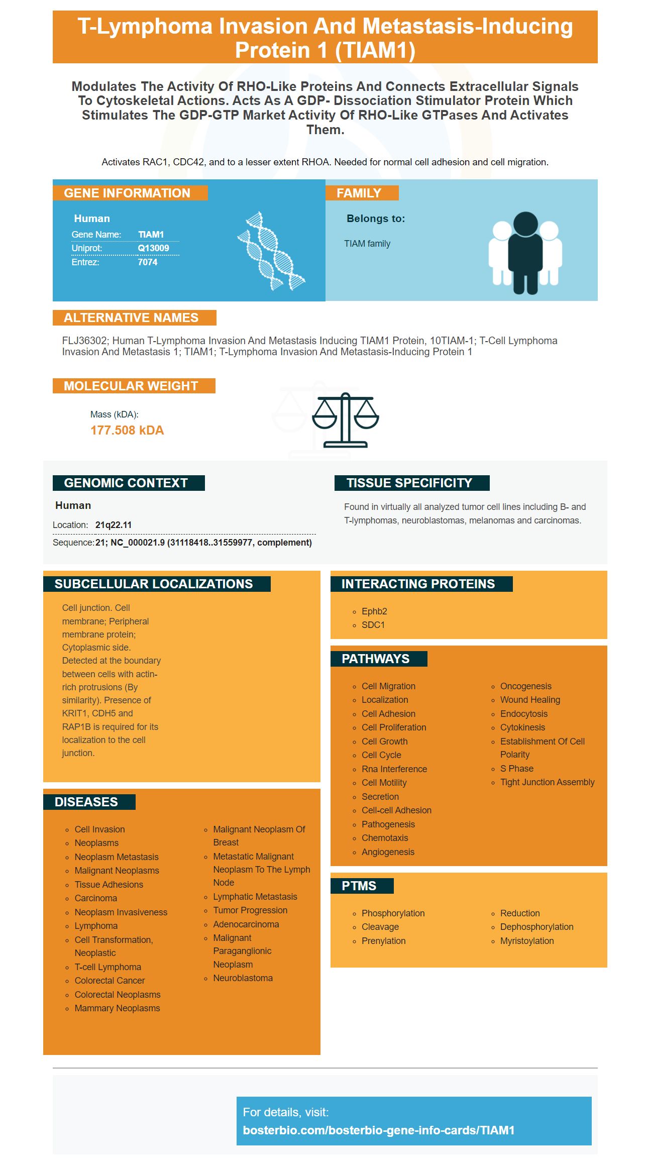
Facts about T-lymphoma invasion and metastasis-inducing protein 1.
Activates RAC1, CDC42, and to a lesser extent RHOA. Needed for normal cell adhesion and cell migration.
Human | |
---|---|
Gene Name: | TIAM1 |
Uniprot: | Q13009 |
Entrez: | 7074 |
Belongs to: |
---|
TIAM family |
FLJ36302; human T-lymphoma invasion and metastasis inducing TIAM1 protein, 10TIAM-1; T-cell lymphoma invasion and metastasis 1; TIAM1; T-lymphoma invasion and metastasis-inducing protein 1
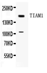
Mass (kDA):
177.508 kDA
Human | |
---|---|
Location: | 21q22.11 |
Sequence: | 21; NC_000021.9 (31118418..31559977, complement) |
Found in virtually all analyzed tumor cell lines including B- and T-lymphomas, neuroblastomas, melanomas and carcinomas.
Cell junction. Cell membrane; Peripheral membrane protein; Cytoplasmic side. Detected at the boundary between cells with actin-rich protrusions (By similarity). Presence of KRIT1, CDH5 and RAP1B is required for its localization to the cell junction.
You've heard of miR-22 , and TIAM1 But do you know how to utilize this gene in your experiments? Did you know that it can affect the growth of tumors and also regulates radioresistance? This article will let you know more about this marker and how you can utilize it in your research. With these guidelines you'll be able to answer to your questions and be able to optimize your experiments.
MiR-22 regulates many mRNAs and has been associated with the progression and development of cancers, including colon cancer. Researchers have studied the role of miR-22 in colon cancer in a new study. The invasion and migration of HCT116 colon cancer cells were inhibited by miR-22 expression. In addition, miR-22 blocked the expression of TIAM1's mRNA as well as its protein which suggests that it is possibly a target gene for miR-22.
Studies in the past have suggested that miR-22 inhibits the expression of TIAM1 through inhibiting its protein expression. The expression of miR-22 in the cells of parents was fourfold higher than in NC cells in this study. Immunocytochemistry experiments confirmed these findings. Furthermore, miR-22 slowed down the colonization capacity of colorectal cancer cells when expressed in nude mice.
MiR-22 prevents the spread of hepatocellular cancer cells by attacking HDAC4 (12). It also inhibits colon cancer cell migration. One study showed that miR-22 expression was suppressive of colon cancer cell invasion. Researchers cultivated HCT-116 cells from their parents with miR-22 in an incubator at 37degC. The researchers then cloned miR-22 precursor sequence of hsamiR-22 into the SD13 expression vector.
MiR-22 was negatively associated with AD patients' levels of inflammatory-cytokines in their serum, and it also inhibited the process of pyroptosis. MiR-22 controlled the expression of gasdermin D and NLRP3 inflammasome, two additional genes implicated in inflammation. In mice, miR-22 lowered GSDMD expression and improved memory.
TIAM1 is a very expressed gene in a variety of types of cancer, particularly metastatic cancer. It can be found in various types of tissues and is especially high in lung and colon cancer. In addition to lung and colon cancer, it is also expressed in the esophagus, nasopharynx, ovary, and pancreas. MiR-22 also inhibits expression of VEGF and Rac.
TIAM1 is a key gene that is found in cancer cells. It is also associated with an invasive nature. This gene is also closely connected to the expression of VEGF, a pro-angiogenic protein. MiR-22 also associates with VEGF which is a signaling molecule which influences cancer cell invasion. Although it is not certain how miR-22 affects cancer, the research findings confirm the role played by this gene in tumor development.
The study also examined the role of miR-22 (GC) by studying the expression levels of mRNA from human fibroblasts. Researchers found that miR-22 directly regulates HuR by binding to its 3'UTR. Researchers also examined the potential roles that miR-22 plays in regulation of VEGF and matrix metallproteinases 2-9 and b-actin.
Additionally, miR-22 negatively affects genes that are part of the ERK signaling pathway genes. MiR-10b binds the 3' UTR Tiam1 of mud crabs and has high compatibility with the Tiam1 3'UTR. With the help of bioinformatics, we identified the genes that are targeted by miR-22. Analyses showed that miR-10b may inhibit the expression of ERK pathway genes in the mud crabs.
The efficacy of radiation therapy is compromised by hypoxic conditions that affect tumors. The metabolic system of tumors is heavily dependent on the accelerated glycolysis, the increased demand for ATP and biosynthetic precursors. The tumor cells are protected from radiation therapy through increased glycolysis, resulting in the formation of radioresistance. The alterations in the glycolytic pathway could be caused by radiation therapy.
FX-11 is one such miRNA. This miRNA regulates radioresistance by promoting DSBs and increasing the cell's ability to apoptose. The miR-34 functions as a tumor suppressor and also blocks LDHA transcription, which enhances the ability of cells to respond to radiotherapy. These results indicate that radioresistance is controlled by the miRNA FX-11.
Acquired radioresistance in various forms of cancer is linked to the overexpression of Aurora A. Recent research has identified Aurora A as a key regulator of radioresistance, which suggests that it could also be a therapeutic target in the treatment of radiotherapy. While this research is preliminary, it will encourage further research into this important regulatory pathway. The effectiveness of radiotherapy could be improved by using radiosensitisers with proven efficacy that inhibit Aurora A.
However, it's not clear what mechanisms are responsible for radioresistance. However, a growing body of evidence suggests that cellular radioresistance is closely associated with the metabolism of tumors. Radioresistance is conferred by the mitochondrial and glycolytic metabolisms of cancer cells. Therefore, targeting these two pathways may help in improving the response of cancer patients to therapies. It could be necessary for the treatment of cancer. This study is an important step towards the development of radiosensitizers which can be used to treat malignant tumors.
For radioresistant pancreatic cancer cells, Wang et al. Two cell lines were developed from pancreatic cancer cells using increasing intensities of IR. The cells were named SW1990-R or PANC-1-R. They were screened for radiosensitivity with the clonogenic survival test. The cells with radiosensitivity had higher IR resistance than the cells of the parent.
In TNBC cells, THOC2 is responsible for stem-like properties and radioresistance by aiding in the release of SOX2 and NANOG from the nucleus. The inhibition of THOC2 and THOC5 also reduces the expression of SOX2 and NANOG which reduces radioresistant TNBC cell growth. Furthermore, reducing the expression of these genes blocks the growth of radioresistant TNBC in the vivo and xenograft tumors.
This study confirms that the expression level of the miR-22 target gene, TIAM1, is an indicator of poor prognosis in colorectal cancer patients. This new information provides new insights into the pathology of colorectal cancer. Researchers also found that miR-22 is a blocker of TIAM1.
A high Tiam1 expression is associated both with lymph node metastasis and shorter overall and disease-free lives. These results suggest that Tiam1 could be a new biomarker to help physicians predict the outcomes of clinical trials. The gene could be used to determine if cancer has metastasized from lymph nodes. But further research is required to understand the significance of Tiam1 expression in tumor metastasis.
TIAM1 can be used in many ways to diagnose and treat different cancers, like lung cancer or melanoma. Its expression level can be used to determine the survival of tumors and the rate of recurrence. It is important to know, however, that TIAM1 has a very limited role in laryngeal squamous-cell carcinoma. This biomarker can be used to identify patients at high risk of recurrence and to personalize surveillance strategies for every patient.
The TIAM1 protein is an element of the Ras superfamily of proteins. It is used as a molecular switch. The TIAM1 protein oscillates between an inactive GTP bound and an active GDP-bound state. TIAM1 is believed to regulate the growth and death of tumor metastasis cells. When the GTP bound form of the protein is elevated, apoptosis-promoting protein is induced.
The study employed an established xenograft mouse model to determine the sensitiveness of LSCC to radiation. In three groups, five mice were injected with either TU686 or Tiam1 cells into the right armpit. The TU686 or Tiam1+ cells were given 10 Gy radiation, or two Gy each, for 8 days after inoculation. The growth of tumors and weight was monitored after 28 days.
This is the first study to demonstrate that Hep-2 cells are more vulnerable to death when they have Tiam1 expressed than in an untreated group. Researchers suggest that the TIAM1 genes could be an effective treatment for a variety of cancers. Researchers are investigating whether TIAM1 could be used to detect cancer patients.
PMID: 7731688 by Habets G.G.M., et al. Sequence of the human invasion-inducing TIAM1 gene, its conservation in evolution and its expression in tumor cell lines of different tissue origin.
PMID: 7753201 by Michiels F., et al. A role for Rac in Tiam1-induced membrane ruffling and invasion.