This website uses cookies to ensure you get the best experience on our website.
- Table of Contents
2 Citations 4 Q&As
2 Citations 16 Q&As
1 Citations 15 Q&As
1 Citations
1 Citations 1 Q&As
Facts about TGF-beta receptor type-2.
The formation of this receptor complex composed of 2 TGFBR1 and 2 TGFBR2 molecules symmetrically bound to the cytokine dimer results in the phosphorylation and the activation of TGFRB1 by the constitutively active TGFBR2. Activated TGFBR1 phosphorylates SMAD2 that dissociates from the receptor and interacts with SMAD4.
Human | |
---|---|
Gene Name: | TGFBR2 |
Uniprot: | P37173 |
Entrez: | 7048 |
Belongs to: |
---|
protein kinase superfamily |
AAT3; EC 2.7.11; EC 2.7.11.30; FAA3; HNPCC6; LDS1B; LDS2B; MFS2; RIIC; TAAD2; tbetaR-II; TGF-beta receptor type II; TGF-beta receptor type IIB; TGF-beta receptor type-2; TGF-beta RII; TGF-beta type II receptor; TGFbetaRII; TGFbeta-RII; TGFBR2; TGF-bRII; TGFR-2; transforming growth factor beta receptor type IIC; transforming growth factor, beta receptor II (70/80kDa) isoform 1; transforming growth factor, beta receptor II (70/80kDa) isoform 2; transforming growth factor, beta receptor II (70/80kDa); transforming growth factor, beta receptor II (70-80kD); Transforming growth factor-beta receptor
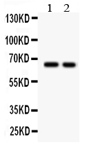
Mass (kDA):
64.568 kDA
Human | |
---|---|
Location: | 3p24.1 |
Sequence: | 3; NC_000003.12 (30606472..30694142) |
Cell membrane; Single-pass type I membrane protein. Membrane raft.
The TGFBR2 marker is a popular biomarker for many biological assays. The TGFBR2 molecule is present in many animal samples, so antibodies developed for this receptor are often monoclonal or polyclonal in nature. Several companies develop antibodies for this marker based on mouse and rabbit samples. In most cases, these antibodies are highly specific and sensitive, allowing researchers to detect the presence of this molecule in biological samples.
One way to detect tumors is to look for the TGFBR2 marker. Mutations in TGFBR2 are associated with increased risk of severe deformity and decreased survival in patients with advanced malignancies. Hypoxia is thought to help maintain cancer stem cells and to support tumor immunity. Furthermore, hypoxia-induced adaptive responses may enhance tumor immunotherapy resistance. The effects of hypoxia on cancer cells may also involve the recruitment of immune-suppressive cell populations and reconstruction of the TME stroma.
In both humans and mice, the TGFBR2 marker is highly correlated with intra-tumoral hypoxia. However, no systematic study has been conducted to assess the relationship between tumor hypoxia and patient outcomes. Therefore, researchers analyzed hypoxia response patterns in bladder cancer and associated clinicopathological and genomic characteristics. They then derived a score called HPXscore, which represents the individual hypoxia-response pattern. A low HPXscore correlated with a low response pattern and high DNA damage repair.
We also analyzed the association of HPXclusterE with high tumor mutation burden. Interestingly, HPXclusterB showed a lower tumor mutation burden than HPXclusterE. Further, we also found that HPXclusterE and HPXclusterB were associated with hypoxia-response phenotype. Furthermore, HPXscore was an independent prognosticator of immunotherapy outcomes. This study suggests that a comprehensive landscape of hypoxia characteristics may aid interpretation of the underlying mechanisms of immune escape and shed light on clinical applications of immune checkpoints and modification therapies.
PI3K/Akt signaling is essential to various viral processes and promotes host response to a wide range of pathogens. In addition to promoting antiviral and proviral activities, this pathway regulates the synthesis of viral proteins. Various retroviruses, DNA viruses, and RNA viruses take over the Akt pathway. To study the role of PI3K and Akt in Boster Bio, read on to learn how it functions and how it helps to combat pathogens.
PI3K/Akt signaling is necessary for cell proliferation and survival. It is activated by the direct or indirect binding of the p85 regulatory subunit to phosphorylated RTK. It then translocates into the nucleus where it promotes transcription of target genes. It also affects the growth and development of adult hippocampal neural progenitor cells and promotes cell division.
PI3K/Akt signaling in a variety of human cancers is facilitated by the pS6 receptor. This protein phosphorylates Akt and leads to an increased level of mTOR. These two pathways control tumor growth, but in the Boster Bio model, they are inhibited independently of one another. In addition to inhibition of mTOR, the PI3K/Akt pathway contributes to the reduction of PDGFRa and PDGFRb expression.
PI3K/Akt signaling is essential for muscle cell viability. Moreover, it maintains skeletal muscle mass and prevents apoptosis. Interestingly, dystrophin-deficient myoblasts show reduced activity and apoptosis. These abnormalities result in altered signaling of the PI3K/Akt pathway, which leads to decreased muscle mass and wasting.
Antibodies to TGF-beta receptor type-2 (TGFBR2) can be purchased in various forms, including monoclonal and polyclonal antibodies. These antibodies have been developed for detecting this protein in a variety of animal samples. Boster Bio uses rabbit and mouse to produce its antibodies. This makes its products more robust and specific. It also offers a range of product credits for sharing results.
The TGFBR2 gene is a key regulator of the PI3K/Akt pathway. Its expression can be inhibited by siRNA. When compared to wild-type TGFBR2 expression, suppression of TGFBR2 significantly reduces PI3K signaling and phosphorylation of downstream markers of the PI3K pathway, including pAkt and p4EBP1.
SOX4 knockdown significantly decreased TGFBR2 expression in HCC1143 cells. The SOX4-V5 gene was inserted in HCC1143 cells by using CRISPR/Cas9 genome editing technology. The gRNA sequence and the altered PAM motif are highlighted in blue and brown, respectively. Western blot and immunofluorescence analysis of SOX4-V5 cells revealed V5 expression in SOX4-V5-cell line. The parental cell did not show any V5 expression.
TGFBR2 is a transmembrane serine/threonine protein kinase. It regulates PI3K/Akt signaling in human basal-like breast cancer cells. Furthermore, TGFBR2 regulates SOX4 activity. Further studies are needed to elucidate the role of SOX4 in TGFb signaling in human tumors.
The expression of the TGFBR2 marker in prostate cancer has been associated with poorer survival rates. The gene is a histone methyltransferase and exerts important oncogenic functions in PCa. High expression of the TGFBR2 marker is associated with poor overall survival and disease-free survival. It is an important molecular target of miR145.
RT-PCR as a use of TGFBR2 markers can identify TGFb signaling in human tumor tissue. This gene has a role in regulating MET, a cancer-suppressor gene. Coregulation of these two genes was detected in breast cancer cell lines and human tumor tissues. However, further studies are needed to confirm this association. In a recent study, a fusion protein, ETV1, was found to increase TGFBR2 expression in human breast cancer cells.
RT-PCR as a use of TGFBR2 markers is a rapid, highly sensitive method for determining the presence of TGFBR2 in human tumor tissues. The TGFBR2 gene is essential for normal skeletal development. When it is deficient, chondrogenesis is inhibited, and cartilage replaces the interzone cells. Furthermore, TGFBR2-deficient mice develop shorter legs and a longer tail.
After E9.5 days, Tgfbr2 was deleted from mouse limb mesenchyme. Transgenic mice were crossed with Cre-mediated recombination under the Prx1 promoter. In addition, Cre-negative mice were used as controls. After E10.5 days, the Tgfbr2 gene expression in mouse limbs was reduced significantly. A functional assay further confirmed that the gene deletion had occurred in the Tgfbr2 marker.
To determine the best use of the TGFBR2 marker, we used a pHAGE-TGFBR2 viral particle to perform overexpression and rescue experiments. The viral particles were then transfected with a control siRNA or SOX4 and cells were harvested 48 hours later for protein analysis. The results were compared to those obtained with the parental cells. Interestingly, the TGFBR2-V5 viral particle showed greater than threefold enrichment relative to the wild-type controls.
In the present study, the SOX4 and SMARCA4 antibodies were used to overexpress the TGFBR2 gene. qRT-PCR was used to assess the interaction between the proteins. In addition, we used the SMARCA4 ChIP-qPCR to identify the enrichment of TGFBR2 at the promoter region in HCC1143 and HCC1954 cells.
Sox4 is a component of TGFBR2, a protein involved in the regulation of PI3K/Akt signaling. It binds to the promoter region of TGFBR2 and forms a complex with SMARCA4. This protein helps activate TGFBR2 expression by regulating downstream PI3K signaling. It also helps regulate resistance mechanisms in TNBC.
TGFBR2 antibodies react with human TGF-beta receptor. Boster Bio Anti-TGF beta Receptor II TGFBR2 Monoclonal Antibody is tested for WB applications. It can be stored at -20°C for up to one year. This antibody reacts with Human, Mouse, and Rat. These species are known to have functional TGFBR2 receptors.
An ELISA for the TGFBR2 markers can determine if tumor cells have an aberrant energy metabolism. This characteristic is a hallmark of many cancer cells and plays a role in the development of colorectal cancer. In addition to this, the marker has other functions, including WNT signaling, matrix re-modelling, and regulating cell migration and cytokinesis.
PCR products containing the TGFBR2 gene were subjected to SSCP analysis to exclude contamination from wild-type TGFBR2. The cells used in the study were DLD1 cells and SW48, both of which harbor homozygous mutations in exon 3. A single human random control was used as a negative control. The PCR product was mixed with 4 ml of loading buffer and placed on ice. Afterwards, the sample was run at 20 mA overnight on a 6% polyacrylamide gel, which contains 10% glycerol.
The pTGFBR2 HCT116 cells secrete fewer exosomes than control HCT116 cells. TGFBR2 determines the cargo of secreted exosomes. These exosomes modulate the local environment of recipient cells. Therefore, TGFBR2 is a potent modulator of exosomal proteins. The ELISA for the TGFBR2 marker accurately measures TGFBR2-dependent exosomal proteins in the recipient cells.
PMID: 1310899 by Lin H.Y., et al. Expression cloning of the TGF-beta type II receptor, a functional transmembrane serine/threonine kinase.
PMID: 7959019 by Nikawa J.; A cDNA encoding the human transforming growth factor beta receptor suppresses the growth defect of a yeast mutant.
*More publications can be found for each product on its corresponding product page