This website uses cookies to ensure you get the best experience on our website.
- Table of Contents
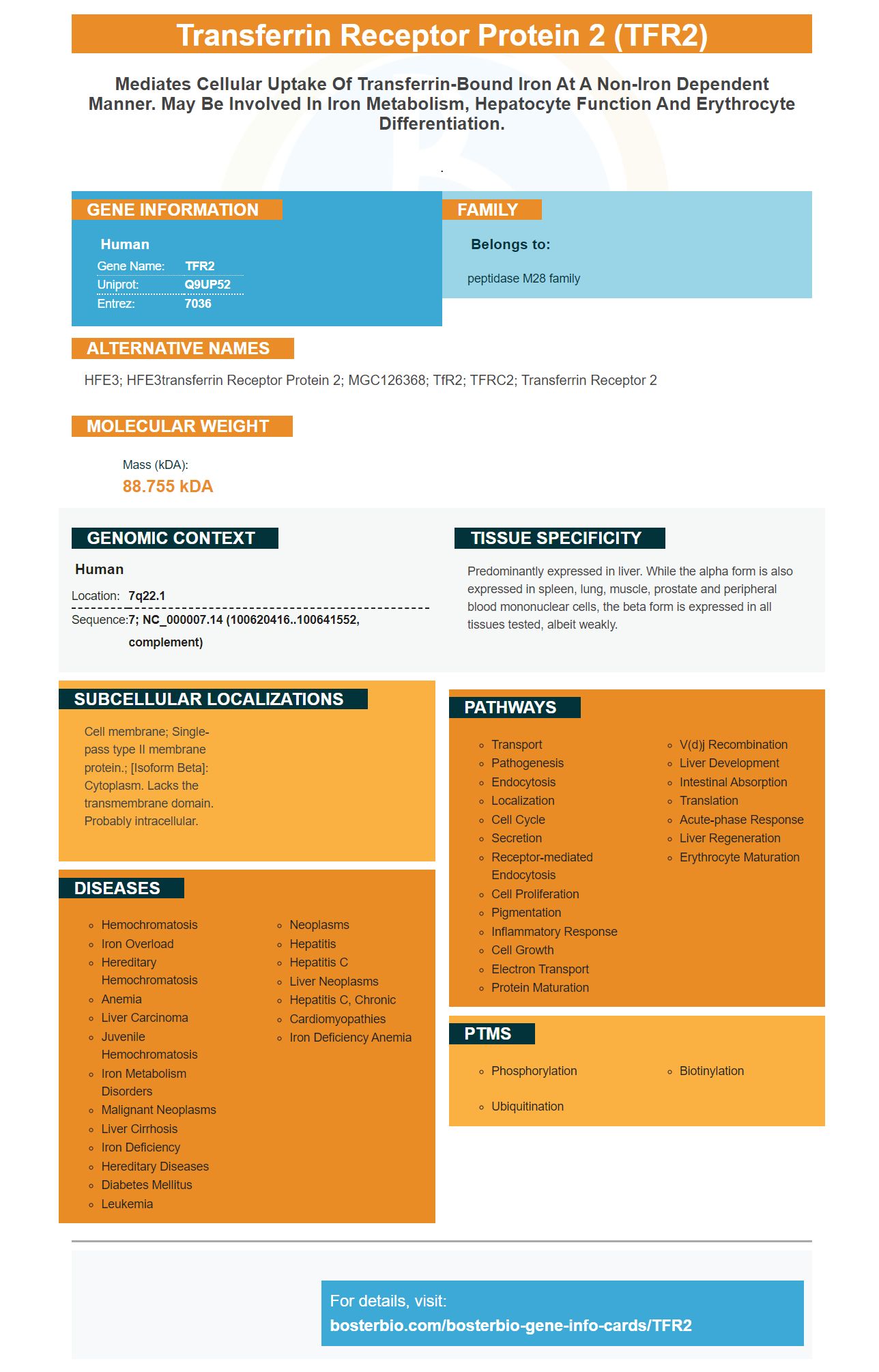
Facts about Transferrin receptor protein 2.
.
Human | |
---|---|
Gene Name: | TFR2 |
Uniprot: | Q9UP52 |
Entrez: | 7036 |
Belongs to: |
---|
peptidase M28 family |
HFE3; HFE3transferrin receptor protein 2; MGC126368; TfR2; TFRC2; transferrin receptor 2
Mass (kDA):
88.755 kDA
Human | |
---|---|
Location: | 7q22.1 |
Sequence: | 7; NC_000007.14 (100620416..100641552, complement) |
Predominantly expressed in liver. While the alpha form is also expressed in spleen, lung, muscle, prostate and peripheral blood mononuclear cells, the beta form is expressed in all tissues tested, albeit weakly.
Cell membrane; Single-pass type II membrane protein.; [Isoform Beta]: Cytoplasm. Lacks the transmembrane domain. Probably intracellular.
In this article, we will discuss the Boster Bio Anti-Transferrin Receptor Protein 2 TFR2 Marker and its uses in Western blot analysis. We will also talk about Steven Boster, his background, and the company’s high-affinity prima antibodies. This article is for scientists in all fields who wish to use this antibody. We will also discuss the advantages of using this antibody for Western blot analysis.
The Boster Bio Anti-Transferrin Receptor Protein 2 TFR2 Marker is a sandwich ELISA kit that detects the protein in Mouse and Rat. This kit reacts with both positive as well as negative samples. It comes in a variety of concentrations. The recommended concentrations should be used to optimize your ELISA test results.
The Online Supplementary Methods describes the procedure for removing membrane and soluble TFR2 cells lysates. For western blot analyses, the total cell or conditioned media is used. A luciferase reporter assay was performed to evaluate the effect of sTFR2 on hepcidin promoter expression.
This antibody detects sTFR2 (a protein involved in iron detection) and TFR1. The antibody can detect these proteins using a variety of methods, including cell culture and basic biochemistry. Boster Bio Anti-Transferrin Repeller Protein 2 TFR2 Marker was used here to detect TFR2 levels in HeLa cells.
The transferrin-binding protein transporter is an essential cell-surface glycoprotein that mediates intracellular iron-binding. The transferrin receptacle regulates iron levels in blood cells by allowing iron to enter the liver cells. The BBB'spermeability is also controlled via the ubiquitous endocytic membrane receptor LRP. The iron in blood is kept from the brain by blocking this receptor. It remains in the liver cells.
One of the most effective routine applications of western blotting is the discovery of protein-protein interactions, and far-western blot is a widely used method for this purpose. It can be used to detect specific proteins, identify post-translational modifications, analyze protein levels, and verify protein expression in cloning programs. There are some common pitfalls with this technique. This paper will provide a detailed explanation of the westernblot protocol as well as its applications in order to avoid them.
Quantitative analysis is difficult because of the presence of weak and uneven target bands as well as the presence membrane background. These problems can be avoided by optimizing upstream steps such as protein loading, membrane cleansing, and blotting-blot preparation. This will allow for brighter target bands. It is important to calibrate the tool correctly and validate its results in quantitative analysis. To do so, titrate proteins that are known in quantity and load blots of protein with known amounts. In other words: the relative quantitation between target bands should be at most two, 1.5, and one.
Normalization is another crucial step in the analysis process. To do a total protein analysis, the proteins must be quantified before being included in normalization calculations. Image Lab Software comes with tools that make this step simple. In addition to assessing total protein levels, one can choose the reference lane using Bio-Rad's Stain-Free technology to minimize the inherent problems with using housekeeping proteins.
A TFR2 marker can be used for protein quantification in western blot analyses. It is highly accurate, versatile, and reliable. One can determine the amount of protein in a sample by subtracting the background from each lanes. Using this marker allows researchers to detect specific protein molecules and identify them in the cells. For the purposes of cell differentiation, this method is especially useful for determining gene expression.
This enzyme in the Western blotting system causes the signal output to peak very quickly, but it also exhausts the substrate rapidly. An optimal system peaks at around 5 min and plateaus for several hours. The optimal system can detect protein at a rate greater than ten times that of enzyme-tosubstrate. It doesn't require the use of darkrooms, which is a major advantage to researchers.
The history of Steve Boster's life is filled with many milestones. Steve has built one of most successful biotech companies worldwide, starting with his early development and birth. Boster was known as "the man who converted science in the bathroom" and has many historical landmarks to his credit. Many of his accomplishments are available online and in books. His biography has been awarded numerous awards. Anyone who is interested about his history should read his biography.
Steve Boster was born in Joliet, Illinois. He died June 26, 2022. Born in Joliet (IL), he was a long-standing sales manager. He served in the U.S. Army as well as being a member of Concordia Hall at Staunton, VA. Boster is survived his 2 Daughters, Crystal Boster & Natosha Peeck, 6 grandchildren, 4 Brothers (Jack & David Boster) and many nieces & nephews.
Boster Bio provides a range high-affinity antibodies to detect TFR2 expression. The company's antibodies have been validated on Western Blotting, Immunohistochemistry, and ELISA. These antibodies are available to researchers at all levels. Boster antibodies are highly sensitive and specific tools for diagnosing TFR2 expression.
Primary antibodies are immunoglobulins, which bind to a specific antigen. Their specificity or affinity will determine their quality. A high affinity antibody will be capable of binding the targeted antigen with minimal binding. Low specificity antibodies will be more able to bind non-targeted antigens. High-affinity antibodies are capable of detection, purification and measurement.
PMID: 10409623 by Kawabata H., et al. Molecular cloning of transferrin receptor 2: a new member of the transferrin receptor-like family.
PMID: 9799793 by Gloeckner G., et al. Large-scale sequencing of two regions in human chromosome 7q22: analysis of 650 kb of genomic sequence around the EPO and CUTL1 loci reveals 17 genes.