This website uses cookies to ensure you get the best experience on our website.
- Table of Contents


Facts about SPARC-like protein 1.

| Human | |
|---|---|
| Gene Name: | SPARCL1 |
| Uniprot: | Q14515 |
| Entrez: | 8404 |

| Belongs to: |
|---|
| SPARC family |

Hevin; High endothelial venule protein; MAST 9; MAST9; PIG33; proliferation-inducing protein 33; SC1; SPARC like 1; SPARCL1; SPARC-like 1 (hevin); SPARC-like 1 (mast9, hevin); SPARC-like 1; SPARC-like protein 1
Mass (kDA):
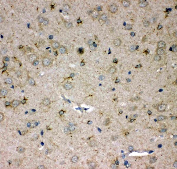
75.208 kDA

| Human | |
|---|---|
| Location: | 4q22.1 |
| Sequence: | 4; NC_000004.12 (87473330..87529503, complement) |
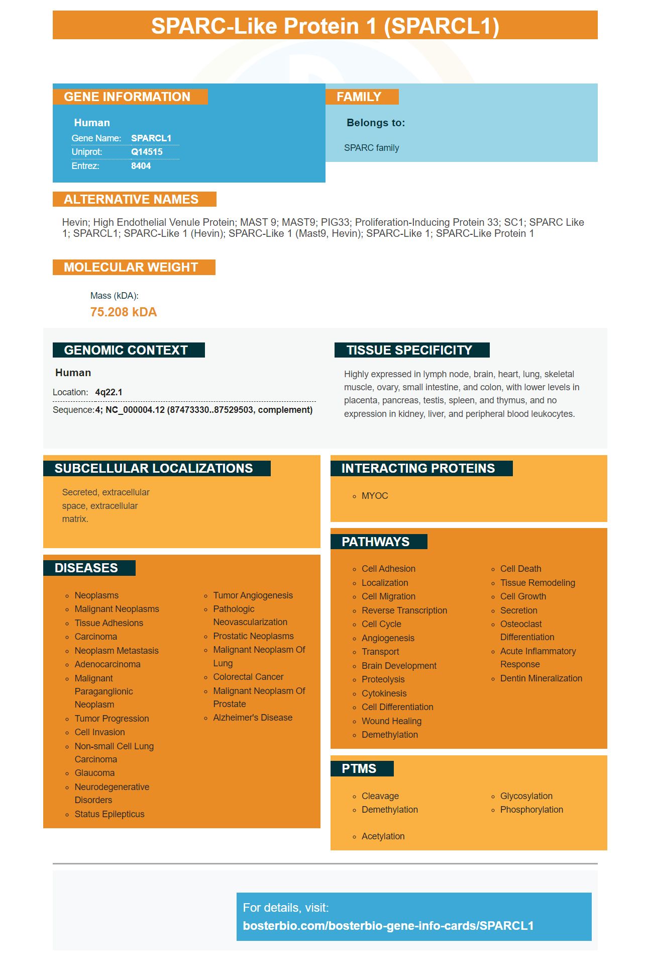
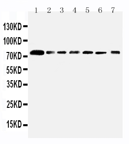
Highly expressed in lymph node, brain, heart, lung, skeletal muscle, ovary, small intestine, and colon, with lower levels in placenta, pancreas, testis, spleen, and thymus, and no expression in kidney, liver, and peripheral blood leukocytes.
Secreted, extracellular space, extracellular matrix.


One of the many features of this antibody is its high specificity and high affinity. Boster validates the antibody on multiple platforms, including with known positive or negative samples. Boster awards product credits to its initial reviewers. Boster is an international company, and is proud to reward scientists from around the world for their contributions to science. You can review this product and other similar products from Boster Bio.
Scientists have recently discovered a biomarker, which can predict Alzheimer's risk. Plasma neurofilament lighting, or PNF is found in the bloodstream of 2,144 Colombians between eight and 75. This test has been used to identify mutation carriers and non-carriers from as young as 22. Rapidly decreasing cognition and shrinking brain cell numbers have been linked to a rapid increase in plasma neuralfilament light.
Researchers are gaining further insight into the brain structure to better interpret amyloid scans. This information can be used to identify a specific stage of a disease. Niklas Mattsson and Oskar Hansson of Lund University used longitudinal data from 741 participants in the Alzheimer Disease Neuroimaging Initiative. They identified four stages for amyloid accumulation. The researchers concluded that the plasma Ab42/40 ratio had a strong correlation with positive PET scans for amyloid deposits in the brain. People in stage zero were not at high risk of developing plaques.
The data came from five thousand brains taken from the ADGC, an academic consortium that includes Banner Alzheimer's Institute and Phoenix. These samples were matched with 989 unaffected donors and autopsy-confirmed Alzheimer's cases. The International Genomics of Alzheimer's Project data was also used. The study results showed that SPARCL1 was able to predict the risk of developing Alzheimer’s disease by 85 percent.
MicroRNA miR-539-3p promotes epithalovarian Cancer by targeting SPARCL1. The gene is responsible for regulating chromosome 7p. It is also known to inhibit expression of CDK14/SPARCL1, which are essential for cell growth. SP1 is the target of miR-539-3p.
This study shows that the microRNA miR-539-3p is a direct target of SPARCL1 and promotes epithal-ovarian cancer progression. This microRNA is negative correlated with advanced clinical stage and lymph node metastasis, and it is downregulated during colorectal cancer. Furthermore, the miR-539-3p/SPARCL1 axis was established using qRT-PCR, scratch wound assay, and cell counting kit-8.
In this study MiR-539-3p promoted E/N-cad, and MiR-539-3p inhibited Snail. MiR-539 in contrast blocked the effects SP1-Snail in PCa cell cells. This suggests that miR-539 might be involved in PCa formation. Different cell lines had different levels of miR-539 expression, which suggests that they could have different effects on migration, invasion, proliferation.
MiR-539-3p targets CDK14 to inhibit cell proliferation. Western blot analysis of two different cell types showed a reduction in the levels of CDK14 relative to the control group. The miR-539-3p and CDK14 combined significantly reduced Ki67 and PCNA expression in ovarian cancer cells. The miR-539-3p/CDK14 combination inhibited cell growth but not apoptosis.
This study also implicated miR-331-3p in suppressing cell proliferation in SKOV3 and A2780 ovarian cancer cell lines. Both cell lines were stopped by miR-3331-3p. The results of the transwell assays showed that miR-331-3p suppresses the migration and invasion of epithelial ovarian cancer cells. However, it is not clear what the exact mechanism may be.
It can be difficult to tell the difference between early and advanced ovarian cancers. Early symptoms are important. Early detection of cancer may be possible by inhibiting the activity miR-539-3p. Treatments targeting this miR might improve patient survival. Patients suffering from ovarian carcinoma could have their outcome improved by inhibiting the enzyme's activity.
MicroRNA-331-3p suppresses RCC2 expression. It inhibits cell proliferation by targeting RCC2, SPARCL1, and RCC2. MiR-331-3p also suppresses tumor metastasis using ovarian cell lines. These studies also show the importance miR-539-3p's role in the prevention and treatment epithelial-ovarian cancer.
It is still not clear how miR-539-3p contributes to the promotion of ovarian carcinoma. The miR promotes EMT by targeting SPARCL1. It targets several genes that are involved in ovarian carcinoma. Many of them are induced when miR-539-3p is absent. MiR-539-3p promotes epithelial ovarian cancer by targeting SPARCL1.
To further investigate the effect of miR-539-3p on the expression of SPARCL1, a new lentivirus containing miR-539-3p mimic was developed. The lentivirus allowed EOC cell-clone transfection with the miR320a/RT/NC vector. qRT-PCR was used to analyze the lentiviruses. After transfection, expression of miR-320a and SPARCL1 was quantified by qRT-PCR.
Researchers compared miR-539-3p protein expression in normal and EOC tissue. EOC tumors were significantly more likely to express miR320a than the adjacent normal ovarian cells. The study revealed that miR-539-3p promotes epithelial-ovarian cancer by targeting SPARCL1 to mice. This study suggests that the miR can be used as a diagnostic tool for ovarian cancer and could even be applied to clinical treatment.
The miR-539-3p copy was co-transfected into 293T oocytes. The cells were collected 48 hours after transfection. They were stained with Renillaluciferase for control. The relative fluorescence of these cells was normalized against Renilla luciferase activity. This result shows that miR-539-3p promotes epithelial and ovarian cancer by targeting SPARCL1.
Western blotting was used to determine the expression levels of proteins involved in epithelial-mesenchymal transition. ASC cells were separated by 10% SDS/PAGE. The proteins were then transferred on to nitrocellulose membranes. To block membranes, 5% milk skimmed was used. The levels of these proteins were determined by using antibodies against E-cadherin Snail and SP1 in ASC samples.
In SKOV3 cells, miR-539-3p was found responsible for controlling SPARCL1 expression. It upregulated the expression of the mesenchymal cell marker vimentin in these cells, which suggests that it may play a role in EMT. Although the mechanism behind EOC is not known, further research will be needed to uncover its molecular basis.
PMID: 7600298 by Girard J.-P., et al. Cloning from purified high endothelial venule cells of hevin, a close relative of the antiadhesive extracellular matrix protein SPARC.


