This website uses cookies to ensure you get the best experience on our website.
- Table of Contents
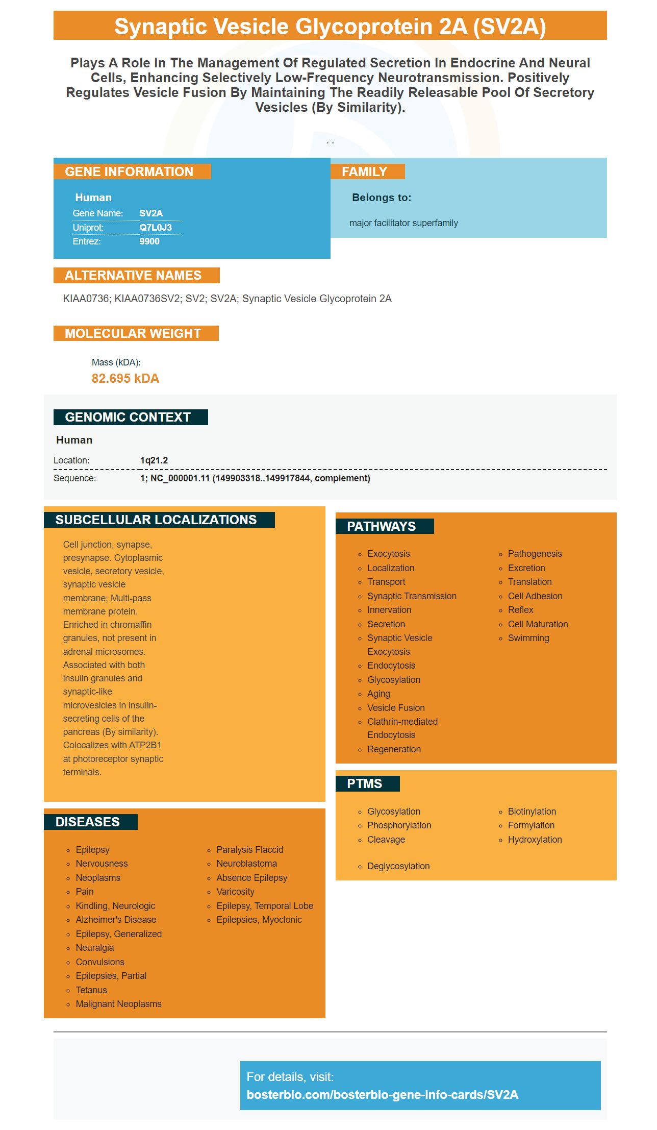
Facts about Synaptic vesicle glycoprotein 2A.

. .
| Human | |
|---|---|
| Gene Name: | SV2A |
| Uniprot: | Q7L0J3 |
| Entrez: | 9900 |

| Belongs to: |
|---|
| major facilitator superfamily |

KIAA0736; KIAA0736SV2; SV2; SV2A; synaptic vesicle glycoprotein 2A
Mass (kDA):
82.695 kDA

| Human | |
|---|---|
| Location: | 1q21.2 |
| Sequence: | 1; NC_000001.11 (149903318..149917844, complement) |
Cell junction, synapse, presynapse. Cytoplasmic vesicle, secretory vesicle, synaptic vesicle membrane; Multi-pass membrane protein. Enriched in chromaffin granules, not present in adrenal microsomes. Associated with both insulin granules and synaptic-like microvesicles in insulin-secreting cells of the pancreas (By similarity). Colocalizes with ATP2B1 at photoreceptor synaptic terminals.


SV2A can be found in nearly all synaptic vesicles as a proton–coupled symporter. The SV2A PCR amplification has identified the biomarker and is now considered a measure of synaptic denseness. However, SV2A has important questions. Its function remains unclear, and its expression is ubiquitous. Although SV2A and synapses are not yet understood, the protein is thought to be associated with synapses primarily made of glutamatergic neurotransmitters.
The researchers speculated that SV2A could function as a biomarker of synaptic density because it is related with neurotransmitter binding. They examined a subset of interneurons in TLE patients, and found that the expression of SV2A was lower in this subset than in other neuronal subtypes. Despite the small number of samples, statistically significant differences were not found between TLE patients versus postmortem controls.
SV2A in hippocampal Excitatory Neurons is co-expressed. Loss of SV2A drives the phenotypes towards convusions. Hippocampal sclerosis is associated to decreased SV2A transcription. However, further research is needed in order to fully understand the biological function of SV2A in synaptic density.
Although SV2A may be a biomarker to synaptic density and a biomarker, its role as a brain disease marker is not well understood. Most of its attention has been directed at targeting SV2A, and selective radioligands for synaptic density measurement have been developed. The changes in SV2A's binding may not predict changes in synapses. Changes in SV2A binding could be due to changes within the protein's binding properties. This in turn may reflect gene expression.
This study used a fusion of two types of imaging techniques to identify SV2A as a biomarker of synaptic densities in brain regions. The cerebellar reference area was used to calculate the DVR. In order to evaluate correlations between global Ab binding and SV2A deposition in broad brain regions, exploratory analyses were also performed by the researchers.
SV2A is a member of the inhibitor of DNA binding-2 (IDB2) family of GABAergic interneurons. It is located within the L1-3 zone. SV2A can also be used as a biomarker for synaptic densities in Boster Bio
The SV2A smporter functions in yeast cells as a proton -coupled smporter. It contributes to the low cytosolic h+ concentration. It may play a role in the co-ordination of galactose with protons, as well as maintaining the pH of neurons. It may also play a role in the synapse-specific uptake of extracellular galactose.
Electrophysiological studies on neurons have shown that SV2A plays a key role in regulating GABAergic Neurotransmission. In the CA3 region of the hippocampus, loss of SV2A reduced action potential-dependent GABAergic neurotransmission, whereas loss of SV2A did not alter action potential-independent neurotransmission. SV2A's absence did not change the morphology or density of the synapse. These studies indicate that the protein is essential for the SV-exocytosis procedure.
SV2A might also trigger synaptic releasing. These two proteins function in parallel. SV2A absence causes Syt1 to recycle more efficiently than Stn2. SV2A Recycling is also increased when Syt1 becomes overexpressed in neurons.
SV2A is involved with calcium-induced exocytosis. SV2A's binding to synaptotagmin with SV2 regulates that activity. Moreover, disruption of synaptotagmin I inhibits SV2 binding, resulting in action potential-independent exocytosis. It seems that SV2 is a neuroprotective function in the brain.
LEV interacts with SV2A, a protein involved in endocytosis. LEV inhibits SV2A SV2A Synaptotagmin Binding and Synaptotagmin Trafficking. Synaptotagmin levels decrease in SV2A -overexpressed synapses. LEV has an impact on synaptotagmin trading and synaptotagmin recycled.
SV2A is found in all parts of the body. However, homozygous mutations account for only one-quarter the live births. It is essential for normal function and embryonic growth. The SV2A-knockout protein makes the pups dependent on insulin and energy metabolism. Management of seizures does not prevent or reverse the growth defect in SV2A-knockout animals.
It has been suggested that SV2A may be involved in the pathophysiology other neurological conditions. Interestingly, SV2A can be found in striatal nurites near neuritic plaques in Alzheimer's disease, but not amyloid deposits, tangles or diffuse plaques. It has been also shown that SV2A significantly expresses in the mitochondria, another parameter that contributes towards AD progression.
The SV2A/-C markers are found in a majority synaptic vesicles. They indicate that they are involved hormone secretion. Both primary chromaffin cell synaptic-like cells and synaptic SV2A/C are home to the SV2A/C markers. Although they have very similar sequences, it is not clear if they play any functional role.
SV2A also exists in the plasma membrane. It is involved the glucose-dependent translocations of granules onto the plasma membrane. SV2A is not required for this process but is necessary to facilitate it. It is also required for the maturation of granules in the synapses. The SV2A marker is present in nearly all synaptic vesicles, but it does not play a significant role in regulating neuronal physiology.
SV2A belongs to the SV2A family which includes SV2B, SV2C and SV2A. The three members in the SV2 Family have 60% sequence homology. They are found within synaptic vesicles. SV2A, the only member in the family, is present everywhere in the adult brain. SV2B & SV2C can only be found at neuromuscular junctions.
In addition to being present in almost all synaptic vesicles, SV2A has also been linked to the inhibitory system. GABAergic terminals have SV2A expressed in over 90%-100% of SV2A expressing principal cell layers. Glutamatergic cells express only 40-50% SV2A+ synaptic vesicles, in contrast to glutamatergic ones. SV2A & VGAT colocalize.
Interestingly, despite the fact that SV2A is present in most synaptic vesicles there are very few studies that have investigated its function. In addition, SV2A immunoreactivity has little effect on the distribution of insulin granules in the NR group, while its absence in CA3 rad layer has no significant effect. These results support the hypothesis that SV2A can be found in almost all synaptic vesicles.
Although the SV2A proteins are thought to be a biomarker for synaptic densit, it is not clear what their role is. The protein is abundant in synapses but its association with them is stronger for glutamatergic and GABArgic terminals. SV2A could be used for neuroimaging studies.
The PCR amplification process may produce a variety results. Boster Bio SV2A is one example of such a result. Primers that anneal at 5' termini are used in PCR amplification. The PCR products are purified using a Wizard SV gel and PCR clean-up system, both manufactured by Promega. Primers 1 and 4, which fuse the genes through overlap extension PCR, are combined. The overlap extensionPCR product is digested in NdeI, BamHI, then ligated using pET-9a .
PMID: 15210974 by Lynch B.A., et al. The synaptic vesicle protein SV2A is the binding site for the antiepileptic drug levetiracetam.
PMID: 29649119 by Gustafsson R., et al. Crystal structure of botulinum neurotoxin A2 in complex with the human protein receptor SV2C reveals plasticity in receptor binding.


