This website uses cookies to ensure you get the best experience on our website.
- Table of Contents
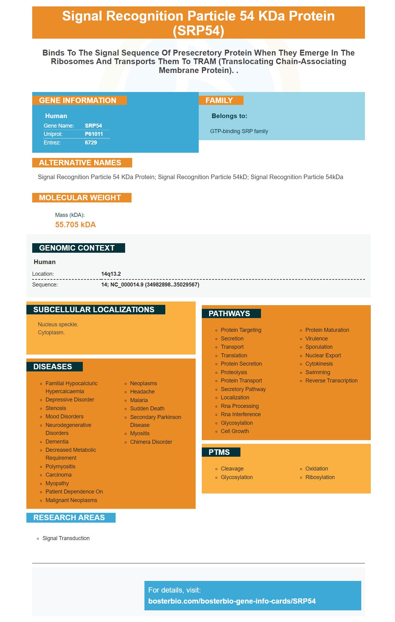
Facts about Signal recognition particle 54 kDa protein.
Human | |
---|---|
Gene Name: | SRP54 |
Uniprot: | P61011 |
Entrez: | 6729 |
Belongs to: |
---|
GTP-binding SRP family |
signal recognition particle 54 kDa protein; signal recognition particle 54kD; signal recognition particle 54kDa
Mass (kDA):
55.705 kDA
Human | |
---|---|
Location: | 14q13.2 |
Sequence: | 14; NC_000014.9 (34982898..35029567) |
Nucleus speckle. Cytoplasm.
The SRP54 Marker is a widely used molecular biomarker for the detection of human fibroblasts. Boster Bio has several high-affinity prima antibodies. These products can be used to measure the efficiency of protein translocation from membranes to cells. Boster's SRP54 markers are available for many applications, including cancer diagnostics, immunohistochemistry, and lipidomics.
This highly specific antibody, first described in 2001, is now the gold standard in SRP54 detection. The antigen is identified by the SRP54 marker. It is present in human tissues. Boster’s high-affinity primary antibodies use this marker for SRP antibody detection. We used the SRP54 marker in a recent study to detect SRP antibodies in serum samples taken from patients with AIDS.
Primary antibodies are immunoglobulins capable of binding to a specific target antibody. Primary antibodies are defined by their affinity and specificity, both of which are dependent on the antigen. High-affinity primary antibodies have high specificity but low affinity. These antibodies are then used for detection, purification, and measurement. The SRP54 antibody is the most widely used for detection in biobanks as well as clinical laboratories.
ELISA is another method to measure specific analytes from crude preparations. High-affinity antibodies are able to bind to epitopes within fixed tissue and wash out non-specific material. This makes ELISA a powerful tool for measuring specific analytes in crude preparations. Because the SRP54 marker is used in this method, the antibodies are stable and can be used to detect specific analytes in crude preparations.
Boster’s high-affinity primary human IgG markers anti-human IgG are highly specific and bind the SRP54 epitope. These antibodies also bind with a wide variety of proteins in the body which increases the assay's sensitivity. The SRP54 marker, which is highly sensitive, is well-known. But there are still a number of limitations.
High-affinity primary antibodies are highly sensitive and selective. Boster's antibodies can also be bound to a variety of immunoglobulins including a subset IgG molecules. This makes them ideal for certain diseases and for diagnosing SRP-infected lesion. But the big question is how will researchers identify these marker?
Biotech Source stocks bulk antibody solutions that can be used in diagnostic and research purposes. They also stock Pel-Freez second antibodies. Bioventix is another supplier of monoclonal antibody. Bioventix makes monoclonal, as well as polyclonal, antibodies. Bioventix specializes in sheep monoclonal antibodies. The second antibody inventory includes a wide variety of products, including polyclonal antibodies, immunoprecipitation kit, and cell purification systems.
There are multiple ways to detect the SRP54 marker. It is a monoclonal antibody that binds to the SRP54 protein from rat and mouse. You can purchase the antibody in both conjugated and non-conjugated forms. The signal recognition part (SRP) is made of four proteins from the SRP family and an Alu domain. These four proteins can be found at the surface of many cells. They are involved in binding with the SRP54 marker.
SRP54 is a signal-recognition particle (SRP), a molecule that targets proteins in ER membrane. This multisubunit complex ribonucleoprotein complex binds on ribosomes. The membrane is where the mature ER-targeted protein anchors. SRP54 is found near the exit tunnel of peptides.
The SRP binds with a low affinity to the TM domain of the ribosome. This sequence exposes SRP’s recognition sequence. Get4/5 It has been shown that interaction with ribosomes via SRP-mediated detection and interaction is possible only through the TA sequence. The SRP54 marker was identified as being important in RNA-based cell technology.
SRP54, a marker that enhances the efficiency of protein transport, is a good choice for membrane staining. This marker is compatible to membranes made from nitrocellulose, PVDF, and other materials. It is sensitive to the amount of protein loaded onto a gel. Electrotransfer losses can cause the actual protein amount visible on the blot to be slightly lower. A minigel 8 cm x10 cm x 1 mm can increase sensitivity by 2-5 folds
SRP54 is an organic molecule that forms noncovalent interaction with proteins. This facilitates better visualization of and measurement of protein transport. SRP54 is excellent at membrane staining, making it suitable in membrane-based and electroblotting experiments. It also forms strong and non-covalent interactions that are beneficial for proteins. The marker is also dimensionally stable, which makes it possible to compare staining patterns for different proteins in the same sample.
After incubating the cells with SRP54, the membrane should be washed three times with TBS Wash Buffer to remove any unbound antibody. Allow the secondary antibody to be added and let it work for 30 minutes in the dark. Repeat the process using 100 mM tris*Cl. Allow the membrane to incubate for the desired amount of time.
SRP54 was first used to identify proteins that had been electroblotted from their native gels. These complexes were separated and their components detected by PVDF membrane stained. This process was enhanced and protein transfer efficiency improved by the SRP54 marker. After binding to the target proteins, Coomassie blu peptides were transferred onto membranes. This increased the detection efficiency of small protein molecules.
Autoradiography uses a sample to generate radiation and a photographic emulsion for capturing the radioactivity in the sample. These results are often very precise, and can be used to interpret a wide variety of medical tests. The process was developed around the middle of the 20th century and is still widely employed in the medical sector. Here are some of the advantages and disadvantages of autoradiography.
The simplicity of autoradiography is the first advantage. Autoradiography is the only procedure that requires the use of film, which has a high degree of dimensional stability. Autoradiography can be performed faster and more efficiently than conventional methods. Recording test results on film takes less than one minute and requires minimal equipment. This process is useful in many medical fields, including the development of drugs.
Autoradiography films are chemiluminescent, which means that they detect a chemiluminescent sample. This type is also known for its high signal-to–noise ratio which improves detection accuracy. Autoradiography films are often paired with cassettes that allow for light-tight exposures below -70°C. Advanced emulsion technology preserves background clarity while improving image resolution.
Radiographic films' sensitivity varies depending on their intended use. Radiographic films can detect up to 1 % of light and have a maximum density equivalent to 1%. The film's characteristics and the processing conditions will determine the radiographic film's maximum density. If you have a sensitive film, you should be aware of this sensitivity range.
PMID: 8722571 by Patel S., et al. Sequence of the highly conserved gene encoding the human 54kDa subunit of signal recognition particle.
PMID: 9511762 by Gowda K., et al. Protein SRP54 of human signal recognition particle: cloning, expression, and comparative analysis of functional sites.