This website uses cookies to ensure you get the best experience on our website.
- Table of Contents
1 Citations 1 Q&As
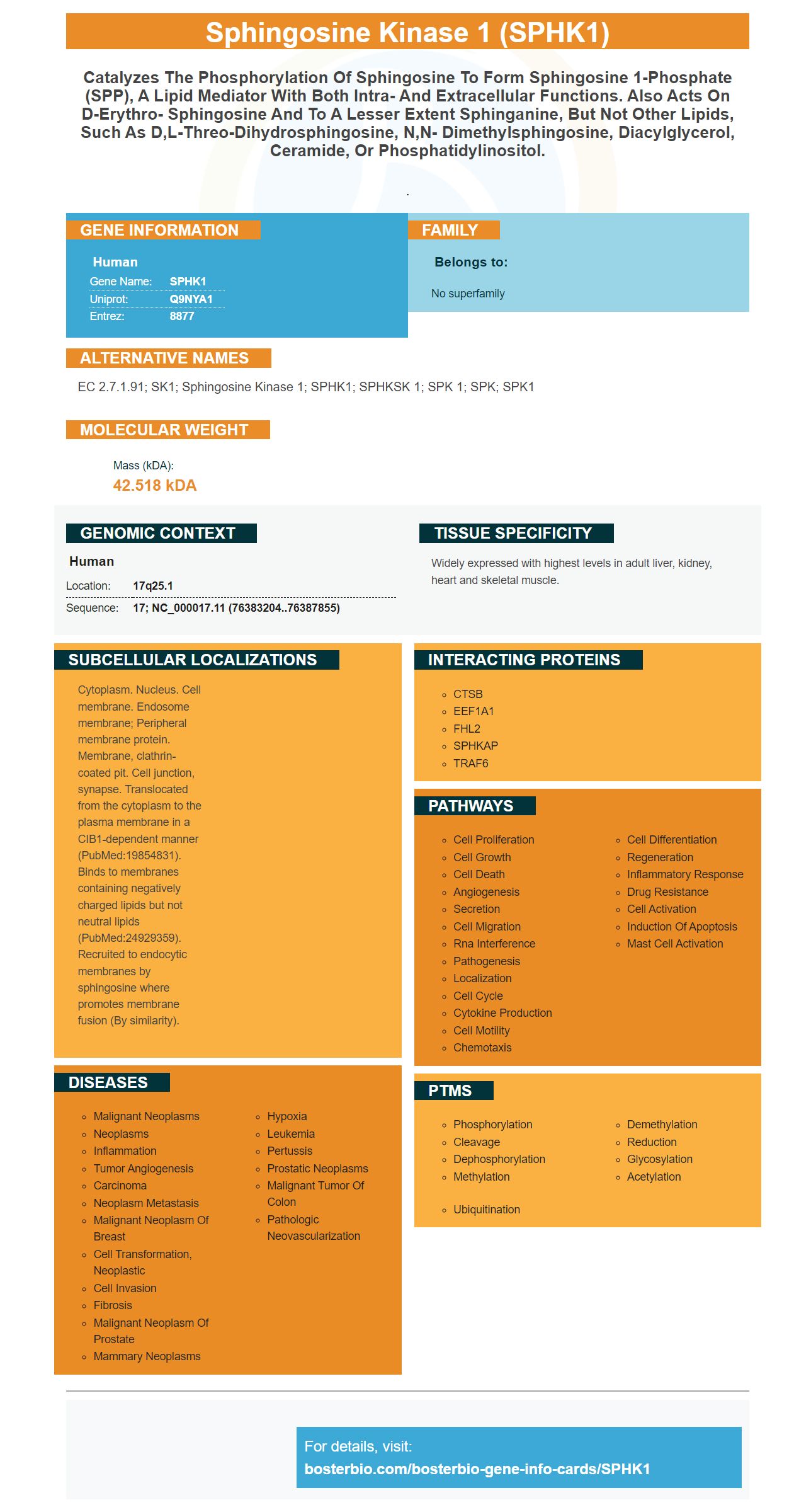
Facts about Sphingosine kinase 1.
.
Human | |
---|---|
Gene Name: | SPHK1 |
Uniprot: | Q9NYA1 |
Entrez: | 8877 |
Belongs to: |
---|
No superfamily |
EC 2.7.1.91; SK1; sphingosine kinase 1; SPHK1; SPHKSK 1; SPK 1; SPK; SPK1
Mass (kDA):
42.518 kDA
Human | |
---|---|
Location: | 17q25.1 |
Sequence: | 17; NC_000017.11 (76383204..76387855) |
Widely expressed with highest levels in adult liver, kidney, heart and skeletal muscle.
Cytoplasm. Nucleus. Cell membrane. Endosome membrane; Peripheral membrane protein. Membrane, clathrin-coated pit. Cell junction, synapse. Translocated from the cytoplasm to the plasma membrane in a CIB1-dependent manner (PubMed:19854831). Binds to membranes containing negatively charged lipids but not neutral lipids (PubMed:24929359). Recruited to endocytic membranes by sphingosine where promotes membrane fusion (By similarity).
You're in the right place if you're unfamiliar with the SPHK1 marker. This article will give you more information about the significance of the SPHK1 marker in autophagy, Tumor vascular signature genes, and S1P receptors. And you'll also discover how to utilize SPHK1 in the lab. Keep an eye out for more details about SPHK1 and other exciting developments in molecular biology research.
For research that tests novel treatments and biomarker discovery, a reliable identification of SPHK1 subtypes is crucial. Evidence suggests that the five primary subtypes of ovarian carcinoma are distinct. SPHK1 expression is linked to optimal tumor debulking, while low-expression SPHK1 is linked to inadequate debulking. These findings highlight the possible significance of SPHK1 in predicting the human ovarian cancer.
In addition to its role in regulating sphingolipid metabolism SPHK1 expression is also associated with low survival for patients with glioblastoma. This gene is associated with poor prognosis. Therefore it is important to discover ways to improve the expression of this protein. Further studies should focus on the role of SPHK1 in chemotherapy-sensitive effects on ovarian cancer patients.
SPHK1 has been proven to increase the growth of tumors and have also been linked to a higher risk of developing cancer. This enzyme plays a crucial function in sphingolipid metabolism, and is a major regulator of the equilibrium of sphingolipids. The overexpression of SPHK1 can lead to inflammation, cancer and tumorigenesis. It improves survival by cutting down S1P levels and inhibiting the production of proapoptotic Sphingolipids.
The SPHK1 gene has been linked to the invading of cancerous esophageal esophag. Exogenous SPHK1 expression increased the invasiveness of EC9706 cells in vitro as well as in mice that were nude. Animal studies revealed that SPHK1 overexpression was associated with increased expression of genes that are involved in the EGFR pathway including EGFR. SPHK1 expression was also linked to the phosphorylation of EGFR.
Research has numerous uses for the SPHK1 marker, which includes the regulation of EMT. It can aid in HCC invasion and metastasis. This marker increases the ubiquitination BECN1 via the TRAF2 protein. These are only some of the many uses for the SPHK1 marker. Below are some additional information. It is recommended that you adhere to the Boster Bio optimization guidelines for the best results.
The SPHK1 protein is required for EMT and is also involved in the degrading of CDh2 by autophagy-linked degradation of lysosomal lysosome. SPHK1 overexpression can promote EMT in HepG2 cell lines. However, it could cause cell migration and invasion to increase when it is excessively expressed. However, when the SPHK1 protein is silenced, the cells are more prone to EMT.
SPHK1 is expressed in the cell membrane of breast cancer cells, and is a marker of the protein. While its precise regulatory mechanism is unknown, it appears to promote autophagy through binding to TRAF2 through lysine 63-linked , ubiquitination. Additionally, SPHK1 induces autophagy in HepG2 cells. The effects of SPHK1 on autophagy are yet to be clarified, so it should be treated with caution.
The SPHK1 marker can be used in a variety of ways and can be used in many experimental settings. It is a good choice for experiments that require cells express SPHK1 both in vivo and vitro. The SPHK1 antiserum inhibits the angiogenic activity of oxLDL human cells. We transfected HepG2 cells using the MYC–SPHK1 gene as well as the corresponding vector to test this hypothesis. To determine the degree of CDh2 expression in the cells, we conducted immunoblotting and fluorescence quantitative RT-PCR.
The SPHK1 protein plays an important role in the angiogenesis of both stromal as well as epithelial cells. The protein is part of the TRAF2 pathway, and its overexpression is linked with the decrease in expression of CDh2 and VIM. We will discuss the most effective applications for SPHK1 in cancer research. Boster Bio: Best Uses Of The SPHK1 Marker
The SPHK1 protein is activated by TRAF2 and BECN1-mediated autophagy and promotes EMT in HepG2 cells. SPHK1 can inhibit migration and invasion when it is expressed too much. This protein is also linked with reduced expression of FN1, VIM and CDH2.
Boster Bio Anti SPHK1 antibody Picoband has been validated for use by humans. The antibody is available for purchase with the request of a validation method and images. Boster Bio's Supervision(tm) method lets you save 30 minutes during IHC and enhance background. Boster Bio's Troubleshooting guides provide useful information to help you solve issues and achieve the most efficient results.
This SPHK1 protein plays a key role in autophagy. Overexpression of it in HepG2 cells increases autophagy. The proteasome is inhibited by MG132 did not affect the degradation of CDh2 in these cells. The protein was not destroyed when chloroquine blocked the lysosome. These results suggest that SPHK1 increases autophagy through lysosomal pathways.
The expression of SPHK1 is increased in tumors originating from the stromal and adipogenic cells, which suggests a connection between SPHK1 with these types of cancers. Furthermore, SPHK1 increases tumor growth and metastasis in mice that are not nude. Furthermore, overexpression this gene accelerated the growth of EC9706 tumor xenografts from mice.
The SPHK1 gene is expressed in tumour vasculature in patients suffering from DLBCL. SPHK1 expression is increased in cancerous tumor cells as well as cancer-associated EC suggesting its involvement in angiogenesis. These cells do not express genes that are negatively associated with SPHK1. This study provides additional evidence for the role of SPHK1 in the development of cancer-related vascular diseases.
The expression of SPHK1 was associated with the invasiveness of cancerous esophageal cell lines. The expression of SPHK1 was determined using immunofluorescence as well as RT-PCR. The highest levels of SPHK1 were observed in EC9706P4 cells. These cells also showed the highest invasiveness. Furthermore, SPHK1 expression was associated with lymph node metastasis as well as the extent of invasion.
In a prior study, SPHK1 protein expression was linked with the outcome of clinical patients with esophageal cancer. For instance, SPHK1 expression was overexpressed in seven of twelve esophageal cancer samples and was not detected in the paired normal epithelia. SPHK1 expression was also higher in 23 cases than normal esophageal tissue.
The IHC applications of SPHK1 are growing in popularity, considering that the protein is an ovarian cancer biomarker. Its increased expression in tumors is associated with enhanced MMP1 promoter activity and the expression of mRNA. Even though SPHK1 expression is not a separate factor from other factors, it is a good candidate for surgical outcome indicators. These findings support the possibility of using this protein as a therapeutic target against cancer.
The SPHK1 gene encodes an SPPK1 polypeptide-kinase that has pro-oncogenic potential. It is commonly overexpressed in solid tumors. In in vitro and in vivo studies, SPHK1 overexpression leads to the formation of tumors. In SCID mice, it is responsible for the growth of cancers. The pro-oncogenic properties of the protein require membrane translocation and phosphorylation.
Proteins that resemble human SPHK1 are available as polyclonal antibody. These are produced by immunizing animals using synthetic peptides that are corresponding to SPHK1. These antibodies are purified by the technique of peptide affinity chromatography. The SPHK1 gene expression in the tumors was determined in these tumors using an IHC-based COSP. There are several uses for SPHK1 in the field of immunohistochemistry, but it is important to understand its biological role.
In vitro studies of endometrioid ovarian cancer, SPHK1 expression has been implicated in a variety of cancer-related processes, including angiogenesis, invasion, as well as metastasis. Additionally, overexpression of SPHK1 has been linked to tumor progression as well as drug resistance. This makes it a possible diagnostic marker for the disease. However, further research is needed to establish the relevance of SPHK1 in clinical settings.
Boster Bio's S1P marker is used in an ELISA to determine the S1P activity of cells. This marker is made by an enzyme called SPHK1. This protein is involved in cell proliferation, migration and angiogenesis. It also plays a role in lymphocyte chemotaxis as well as inflammation. SPHK1 is created in the vivo by the enzyme SPHK1, which is activated by an extracellular receptor agonist. Tumor cells cannot grow or migrate in the event that this enzyme is not inhibited. Zhang et al. Zhang et al. demonstrated that inhibiting SPHK1 expression diminished the migration and proliferation capabilities of the hepatocellular carcinoma cell lines.
SPHK1 activity is measured in cells using the commercial SPHK1 Assay kit. A competitive ELISA kit was used to measure the S1P levels. In addition, the TC (thyroid carcinoma) marker decreased the migration and growth of thyroid cancer cells in a mouse model with NOD/SCID. A decrease in SPHK1 also slowed Notch signaling and cell migration of thyroid cancer cells.
Boster Bio's SpHK1 marker is used in an ELISA to detect specific analytes within basic preparations. The antibodies are able to be washed away when the sample isn't specific to the protein. Therefore, ELISA is a powerful tool to detect specific analytes within simple preparation. In addition to SPHK1, other markers could also be useful.
PMID: 10863092 by Melendez A.J., et al. Human sphingosine kinase: molecular cloning, functional characterization and tissue distribution.
PMID: 10802064 by Nava V.E., et al. Functional characterization of human sphingosine kinase-1.
*More publications can be found for each product on its corresponding product page