This website uses cookies to ensure you get the best experience on our website.
- Table of Contents
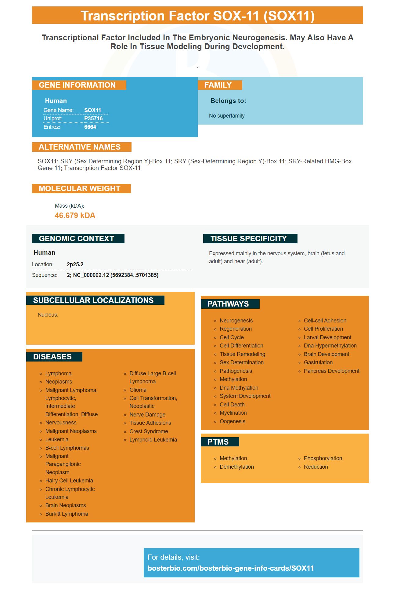
Facts about Transcription factor SOX-11.
.
Human | |
---|---|
Gene Name: | SOX11 |
Uniprot: | P35716 |
Entrez: | 6664 |
Belongs to: |
---|
No superfamily |
SOX11; SRY (sex determining region Y)-box 11; SRY (sex-determining region Y)-box 11; SRY-related HMG-box gene 11; transcription factor SOX-11
Mass (kDA):
46.679 kDA
Human | |
---|---|
Location: | 2p25.2 |
Sequence: | 2; NC_000002.12 (5692384..5701385) |
Expressed mainly in the nervous system, brain (fetus and adult) and hear (adult).
Nucleus.
The SOX11 Marker antigen has a variety of uses in biology. It is used as a diagnostic and prognostic marker for mantle lymphoma cell. SOX11 also promotes osteogenic differentiation and the proliferation of mesenchymal progenitors. It also regulates Wnt7b. Find out more about this antigen and its applications.
In recent times, SOX11 has emerged as a significant transcription factor in the pathogenesis of mantle-cell lymphoma (MCL) even though it is absence in normal B cells or other mature B-cell neoplasms. This review examines the role of SOX11 as a prognostic and diagnostic marker in MCL as well as its new target of transcription and the mechanisms of regulation.
Mantle cell lymphoma has irregular contours of the nuclear nucleus, condensed and inconspicuous nucleoli, scant cell cytoplasm, and condensed chromatin. Blastoid mantle lymphoma, however displays a monotonous growth of tumor cells that are small in size. Pleomorphic mantle cell lymphoma has large cleaved nuclei and distinct nucleoli.
Sox11 is a transcription factor which normally is found in the CNS of embryos. In MCL it is expressed at extremely high levels, and thus serves as a diagnostic and prognostic marker. The levels of expression are highly specific for MCL and have been confirmed to be independent of CCND1 overexpression and t(11;14) translocation. SOX11 expression levels are linked with overall and progression-free survival.
According to the International Prognostic Index, SOX11 levels of mRNA were associated with the ECOG-2 stage, a low risk, and no bone marrow involvement. A positive correlation between SOX11 expression and ECOG-2 stage was found in the survival of patients with MCL CCND1-negative. A statistically significant value was defined as a p value less than 0.05.
In addition to assessing the tumor burden in MCL, SOX11 protein expression is also observed in a number of other lymphoid cancers. Of these, SOX11 protein expression is found in about 30 percent of MCL patients, compared with a low percentage of patients suffering from the same disease. The expression of SOX11 is greater than the expression of cyclin D1 which suggests that SOX11 is a further prognostic factor in MCL.
SOX11 expression levels can also be used as an indicator of prognosis and diagnostics. They are also related to the development of MCL in situ. A significant correlation has been found between SOX11 expression levels and the progression of MCL to disseminated stages. This study suggests that SOX11 expression could be an indicator of prognosis and diagnosis for mantle-cell lymphoma.
The SOX11 gene encodes a nuclear protein. Multiple cell lines including the KM3 acute lymphoblastic leukemia cell line, have been found to express SOX11. The SOX11-C1 reagent can be used to confirm specificity in imaging applications. In cells that express SOX11 exhibit nuclear localization.
SOX11 is a member of the SOX protein family and is a crucial diagnostic and prognostic marker in epithelial ovarian cancer and in mantle lymphoma cells. There are numerous studies that suggest that SOX11-targeting tests do not have specificity and are low in background. One promising monoclonal antibody to SOX11 is SOX11-C1 It has been tested for use in western blots, flow cytometry, and IHC-P.
Sox11 is a vital growth regulator in neoplasms. Therefore the development of antibodies against SOX11 must be developed for experimental use. The SOX11 antibody was created in accordance with the Clinical and Laboratory Standards Institute's guidelines for laboratory workers. SOX11 is a monoclonal, approved antibody, was an important addition to the company's product line.
A recent study using an antibody against SOX11 has found that the SOX11-C1 detects the presence of tumor cells that are circulating in various cell types. It was also found to exhibit strong staining of MCL cells' nuclear membranes and weak immunoreactivity in the JEKO-1 and GRANTA-519 cell lines. Similar to SOX11-C1, SOX11-C1 antibody exhibits specific staining of SOX11-positive cells within WB.
100% of cases containing MCL were analyzed by the SOX11-C1 antibody. Other tissues showed moderate SOX11 staining. This antibody was used to reclassify cases that were previously believed to be negative for SOX11 due to low SOX11 levels. The SOX11-C1 antibody in addition to its prognostic function was negative for a range of HCL cases, with the exception of Burkitt's lymphoma.
SOX11-C1 antibody, which recognizes the SOX11 gene is used as loading control. This antibody can be used to quantify protein, which can help determine whether the SOX11 gene in an HLA+ positive cell type is functional. The data analysis was carried out with FACS Canto II. One gram of the SOX11-C1 antibody was used for each cell line.
The cytokine ICA promotes osteogenic differentiation in mesenchymal stem cells (BMSCs) cells. This molecule is a powerful inducer of bone formation and has many benefits. It acts as a bone-regenerating factor by increasing BMSC proliferative capacity and altering expression of alkaline-phosphatase. It also regulates expression of PDZ binding protein, a transcriptional co-activator.
The effects of hypoxia can also benefit mesenchymal cells by encouraging their proliferation and osteogenic differentiation. It also reduces the process of adipogenesis. Cytokine antibody array analysis of MSCs exposed to hypoxia revealed up-regulation of osteogenic and proliferation-related genes. Thus, adipogenic and osteogenic potentials of mesenchymal stem cells could be due to the different expression of growth factors.
IGF-1 is a major regulator of the bone formation process. This gene is accountable for helping promote the proliferation and osteogenic differentiation mesenchymal stem cells. It is crucial for bone growth. However the molecular reason behind its actions isn't evident. A recent study suggests that it can promote bone development in mice by altering the expression of the BMSC protein Wnt3a.
Studies of mesenchymal stem cells have shown that higher levels of extracellular Ca2+ can enhance the capacity of mesenchymal precursor cells to differentiate into osteoblasts that form bone. OPN enhances osteogenic differentiation mouse models, in addition to stimulating bone formation in mice. This study also reveals that OPN is essential for bone remodeling.
IGF-1 in mice can have osteogenic effects on mesenchymal precursor cell proliferative capacity. Apigenin and amentoflavone also aid in osteogenic differentiation. In animal studies, IGF-1 stimulates bone growth by increasing the expression of ALP, Runx2, and OCN genes.
The extracellular calcium present in MSCs facilitates the migration process, and elevated levels of OPNs cause the expression of osteogenic genes, such as OPNs. This is an essential step in the process of osteogenic differentiation. The protein is needed to produce osteoblasts. These findings require further investigation. While OPN is a paracrine molecule that promotes osteogenic differentiation and proliferation of mesenchymal progenitor cells
The SOX11 marker regulates WnT7b expression in mouse trabeculae. Wnt7b overexpression can hinder osteoclast differentiation and activity, which results in a decrease in bone mass. This is consistent with the effects of Wnt7b's on osteoclast activity, bone resorption, and osteoclast differentiation. The osteoporosis treatment might benefit from the pharmacological inhibition of Wnt7b.
In BMMs, the overexpression of Wnt7b inhibited osteoclast differentiation through inducing an increase in the mTOR pathway as well as the nuclear factor kappa B (NF-kb) signaling pathway. It also inhibited the phosphorylation of p65which is an essential component of the signaling pathway for NF-kb. The overexpression of Wnt7b inhibited the activation of NFKb at the Ikk-a protein level. However, Wnt7b did not significantly alter the phosphorylation of p38 or JNK.
Wnt signaling plays a crucial role in osteoclast differentiation. It is well-established that Wnt is a key regulator of homeostasis in the skeleton. Wnt ligands affect osteoblasts and osteoclasts in a biphasic way. For example, imposing the dominant-active form of b.catenin results in a higher bone mass phenotype, and impaired osteoclastogenesis.
The osteoclast number in the BMMs of Wnt7bLysM mice models were lower than those in Wnt7b overexpression mice. The intracellular acidification was also lower in Wnt7bLysM's BMMs. Furthermore, qPCR tests revealed that SOX11 repressed the expression of osteoclast-specific genes.
With the help of tissue microarrays and entire tissue sections, we examined the expression of SOX11 protein. The SOX11 immunohistochemical analysis revealed that eight of nine MCL showed high SOX11 levels of protein. However, SOX11 expression was identical in two of three B-cell lymphomas. However, SOX11 was undetectable in other types of lymphomas.
Wnt7b reduces the canonical Wnt pathway in BMMs. Our data show that SOX11 regulates Wnt7b transcription in the lungs of mice. This demonstrates the importance of SOX11 for the Wnt pathway. We hope that this study will clarify the way in which BMMs are formed in mice. And if we are able to find a suitable treatment for lung cancer, it will help patients.
PMID: 8666406 by Jay P., et al. The human SOX11 gene: cloning, chromosomal assignment and tissue expression.
PMID: 10574465 by Azuma T., et al. Human SOX11, an upregulated gene during the neural differentiation, has a long 3' untranslated region.