This website uses cookies to ensure you get the best experience on our website.
- Table of Contents
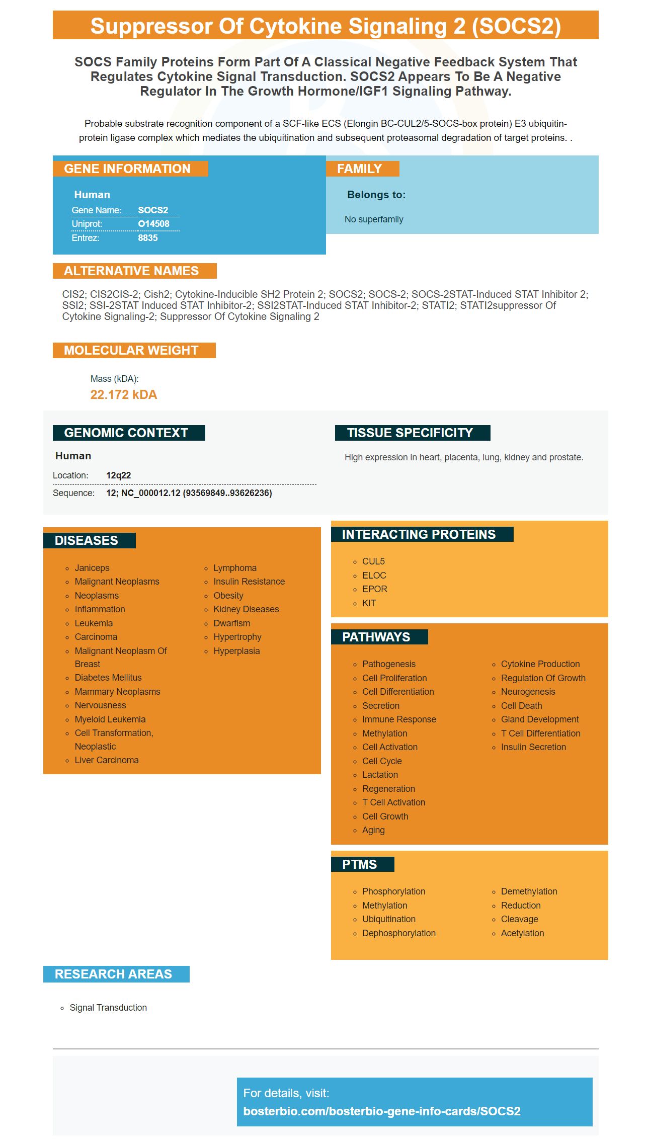
Facts about Suppressor of cytokine signaling 2.
Probable substrate recognition component of a SCF-like ECS (Elongin BC-CUL2/5-SOCS-box protein) E3 ubiquitin- protein ligase complex which mediates the ubiquitination and subsequent proteasomal degradation of target proteins. .
Human | |
---|---|
Gene Name: | SOCS2 |
Uniprot: | O14508 |
Entrez: | 8835 |
Belongs to: |
---|
No superfamily |
CIS2; CIS2CIS-2; Cish2; Cytokine-inducible SH2 protein 2; SOCS2; SOCS-2; SOCS-2STAT-induced STAT inhibitor 2; SSI2; SSI-2STAT induced STAT inhibitor-2; SSI2STAT-induced STAT inhibitor-2; STATI2; STATI2suppressor of cytokine signaling-2; suppressor of cytokine signaling 2
Mass (kDA):
22.172 kDA
Human | |
---|---|
Location: | 12q22 |
Sequence: | 12; NC_000012.12 (93569849..93626236) |
High expression in heart, placenta, lung, kidney and prostate.
In this article, we'll review the functions of SOCS2 and how it relates to apoptotic pathways. We'll also discuss its negative effects on mitochondrial fatty acids oxidation and its role as IGF-1 growth control. We will then discuss how scientists around world can benefit by using the SOCS2 markers in their research.
CYCS, or caspase-9 allows activation of caspase3, a protein that triggers apoptosis. Caspase 9 then cleaves Bid, which leads to cell death. Several studies have been done on the role CYCS plays in apoptosis. More research is needed to determine its role.
CYCS is an essential component of an endocytosis-related protein called Cep-1. It plays a crucial role in germ cell apoptosis and DNA damage-induced apoptosis. It is also essential for tissue regeneration and remodeling throughout the life span. In the following sections, we discuss the role of CYCS in the process. We will then discuss its role in apoptosis.
Cycs, a small hemoprotein, is involved in the electron transportation chain. CYCS is activated and activated by ligands, such as Fas, when apoptotic circumstances arise. Moreover, CYCS also plays a key role in the intrinsic apoptotic pathway. The enzyme is involved in activation of caspase-9 by binding to a death receptor, APAF-1.
Cytochrome c is a heme protein that is located within the mitochondria of aerobic cells. It is involved in electron transportation and has a mass of 11,458. Cytochrome c, which is released by mitochondria, is thought to be part of an apoptotic pathway to respond to DNA damage. Cytochrome c may also play an important role in different apoptotic signaling cascades.
Besides regulating CEP-1 abundance, CYCS also plays a pivotal role in preventing apoptosis triggered by double strand breaks. Double strand break naturally occurs during meiotic-recombination. Incomplete recombination repair could cause apoptosis for late pachytene cell. This suggests that CEP-1 plays an important role in the apoptotic pathway.
Mutations in CYCS inhibit the apoptotic pathway. Mutations in clk-2 suppress DNA damage response phenotypes, including lethality, cell cycle arrest, and DNA repair failure. Mutations in ung-1 inhibit clk-2, which may have a role in base excision repair. Apoptosis is an important part of disease progression in cancer.
SOCS2 is a member the SOCS protein family that also includes SOCS1–SOCS7. Its N-terminus is variable in length, and it has a central SH2-domain and a highly conserved C-terminal SOCS-box motif. Its PDB entry is 2C9W. SOCS2 expression has been shown to be upregulated by mice in the adipose, which helps prevent excessive lipid accumulation in these tissues. SOCS2 is involved in abdominal fat deposition in both SLD and GHR mutations.
SOCS2 can be described as a multifunctional protein, with multiple functions in the nervous system. It regulates nerve growth factor signaling by binding to tropomyosin receptor kinase A (TrkA). This protein is an essential component of neural progenitor neurons and promotes differentiation into them. SOCS2 also blocks GH mediated downregulation of neurogenin-1(NGN-1) neurons. SOCS2-/ mice display decreased neuronal density, fewer neural stem cells, as well as fewer NGN1 expressing neurons in their cerebral cortex.
SOCS2 decreased fatty acid oxidation in mouse Adipocytes. SOCS2 reduced LepR expression and the oxidation palmitate to CO2 as well as the phosphorylation CPT-1b, ACC and JAK. These results suggest SOCS2 may play a part in preventing fatty Acid Oxidation.
Cish-/ mice showed increased SOCS2 expression and an increase in leptin-induced Socs3 induction. However, compensatory mechanisms may be at work here. In the current study, we have determined that the aforementioned compensatory mechanisms are at work in mice. The data presented here are the result of three independent experiments. Once a protein is confirmed to be leptin receptor mediated adipose, it can be collected.
In the increase in expression of SOCS2, a positive feedback mechanism has been suggested. Leptin inhibits SOCS2's activity. Socs2 inhibits leptin's ability to promote fatty acid oxidation in adipocytes. SOCS2 decreases leptin receptive expression and inhibits CIS Recruitment. SOCS2 also inhibits the recruitment and activity of elongin B/C.
One study concluded that supplement SOCS2 significantly decreases p -ACC and CPT-1b concentrations in mitochondria. It also inhibited cyclooxygenase-2 or AOX1 activity and reduced ATP levels. Forced SOCS2 overexpression was also a factor in preventing mitochondrial fatty acids oxidation.
Another study showed that SOCS2 decreased mitochondrial fatty acids oxidation in mouse Adipocytes compared to control cells. The compound decreased the expressions of p–AMPK/JAK, two enzymes that are crucial in mitochondrial fatty Acid Oxidation. Further, Ad-SOCS2 significantly reduced the phosphorylation of ACC and AMPK. It also reduced the expression rate of the leptin-receptor gene, which is quite interesting.
SOCS2 inhibits fatty acids transporters. However the mechanism that causes the inhibition of mitochondrial fat acid metabolization by the enzyme is still not clear. SOCS2 plays an important role as a suppressor in cytokine signals and in the development, fat deposition, skeletal muscle, and the central nervous system. It is still not clear how SOCS2 regulates mitochondrial fatty acids oxidation. The enzyme is important for fat deposition and metabolism as well as the regulation of inflammation.
It is important that SOCS2 plays a role in fatty acid metabolism. FABP4 is overexpressed, which inhibits fatty acid metabolism and reduces the amount of free fat acids in the mitochondria. It also blocks the Akt/mTOR signaling pathway and prevents leptin-induced mitochondrial fatty acids oxidation.
Numerous researchers have confirmed this effect. Costa, Almeida und Jacobsen studied the effects of antioxidant Boster Bio SOCS2 on mitochondrial acid oxidation. Researchers also found that antioxidants such as vitamin E can have similar effects on liver function. However, BOSTER Bio SOCS2 had a greater effect on the oxidation fatty acids in mice than it did in humans.
The growth hormone/IGF-1 signaling mechanism was a subject of immunology in the past. Recent research has shown it to be a pertinent topic in the field. The GH/IGF-1 axis plays a role in numerous physiological processes and is critical in the development of aging. It is believed that SOCS2 regulates GH/IGF-1 signaling.
SOCS2 plays a key role in GH/IGF-1I signaling. It is also known that it acts as an effector of GH to the skeleton. Studies have shown mice without SOCS2 are more likely to grow and are 40% heavier than their littermates after week three. SoCS2 overexpression inhibits the activation of other SOCS by IGF-1, which in turn inhibits signaling from those genes.
In addition to its role in IGF-1 growth control, SOCS2 also affects the maturation of proinsulins. It has a direct effect on the formation and maturation o PC1, which regulates IGF-1. Although this effect is not compatible with life, mice with higher growth rates are found to have overexpression of SOCS2. Further research is required to determine the role SOCS2 has in IGF-1 signals.
SOCS2 can also be associated with the control IGF-1 signaling of breast cancer cells. This is likely because SOCS2 has a similar effect on SNHG7. So far, the effect of SOCS2 on breast cancer cell proliferation has been shown in mouse models. One study showed siRNA targeting SNHG7 to significantly decrease the number cells, including MCF7 as well as MDA-MB-231. The RNAi treatment also significantly inhibited the proliferation of MCF10A breast cancer cells.
RNAseq analysis also revealed that reduced SNHG7 expression did not prevent IGF from activating the signaling cascade, and decreased the regulation of most IGF1 target genes. RNAseq data also revealed a global correlation in IGF1 induced and SNHG7 knockdown. Additionally, gene sets enriched for IGF1-regulated genes were highly correlated with SOCS2 expression.
PMID: 9266833 by Minamoto S., et al. Cloning and functional analysis of new members of STAT induced STAT inhibitor (SSI) family: SSI-2 and SSI-3.
PMID: 9344848 by Masuhara M., et al. Cloning and characterization of novel CIS family genes.