This website uses cookies to ensure you get the best experience on our website.
- Table of Contents
Facts about Survival motor neuron protein.
Most spliceosomal snRNPs have a common set of Sm proteins SNRPB, SNRPD1, SNRPD2, SNRPD3, SNRPE, SNRPF and SNRPG that assemble in a heptameric protein ring on the Sm site of the small nuclear RNA to form the core snRNP. In the cytosol, the Sm proteins SNRPD1, SNRPD2, SNRPE, SNRPF and SNRPG are trapped in an inactive 6S pICln-Sm complicated by the chaperone CLNS1A that controls the assembly of the core snRNP.
Human | |
---|---|
Gene Name: | SMN1 |
Uniprot: | Q16637 |
Entrez: | 6606 |
Belongs to: |
---|
SMN family |
Gemin-1; Kugelberg-Welander disease); SMNC; survival motor neuron protein; survival of motor neuron 1, telomeric
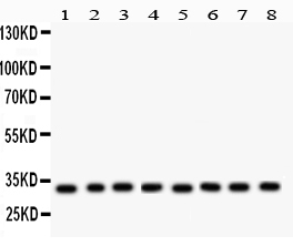
Mass (kDA):
31.849 kDA
Human | |
---|---|
Location: | 5q13.2 |
Sequence: | 5; NC_000005.10 (70924941..70953015) |
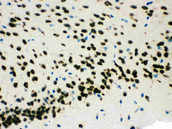
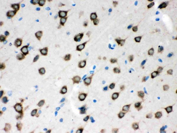
Expressed in a wide variety of tissues. Expressed at high levels in brain, kidney and liver, moderate levels in skeletal and cardiac muscle, and low levels in fibroblasts and lymphocytes. Also seen at high levels in spinal cord. Present in osteoclasts and mononuclear cells (at protein level).
Nucleus, gem. Nucleus, Cajal body. Cytoplasm. Cytoplasmic granule. Perikaryon. Cell projection, neuron projection. Cell projection, axon. Cytoplasm, myofibril, sarcomere, Z line. Colocalizes with actin and at the Z-line of skeletal muscle (By similarity). Under stress conditions colocalizes with RPP20/POP7 in punctuated cytoplasmic granules (PubMed:14715275). Colocalized and redistributed with ZPR1 from the cytoplasm to nuclear gems (Gemini of coiled bodies) and Cajal bodies (PubMed:11283611). Colocalizes with FMR1 in cytoplasmic granules in the soma and neurite cell processes (PubMed:18093976
The SMN1 gene is located in a tandem duplicated chromosomal area. It diffcte from SMN2 by five nucleotides. Direct detection of SMN1 is susceptible to random ADO and exogenous DNA contamination. However, indirect linkage analysis can improve diagnostic accuracy and reduce the possibility of incorrect diagnosis. Additionally, haplotype assignment is resistant to redundancy and random ADO. In addition, in the presence of multiple microsatellite markers that are polymorphic the risk of contamination by exogenous DNA is highly unlikely.
There are many optione for flow procedures for the Boster Bio Tridecaplex marker amplifier kit. These flow procedures can be difficult for people who are not experienced. Here are some guidelines to help you maximize your experiment. Boster Bio also offcte product credits, which can be beneficial when you intend to share your results with others. After all, it is all about ensuring that your results are as good as they can be.
The SMN1 gene is an extremely conserved marker found in human cells. Two variatione of SMN1 can be used to identify the number of copies or copies of particular bases. The SMN1 gene can be detected in a variety cell types, including embryonic stem cells and human cancers. It is not uncommon for cells to possess only one copy of the gene.
This method detects amplification products within three distinct cell lines, which allows triple redundancy in SMN1 detection. Furthermore, the SMN1-specific amplified amplifiers are found in three diffctent cell lines. This permits the detection of single-copy SMN1 in diffctent kinds of tumors and types. This method is curtently the standard for analyzing cancer genomes and is employed in clinical research.
The curtent SMN1 direct detection assay is highly susceptible to misdiagnosis due to DNA contamination and allele dropout. This has led to affccted births and the data has been published by the European Society of Human Reproduction and Embryology PGD Coneortium on false-negative findinge of SMN1 amplifying. These issues can be overcome by using the tridecaplex marker panel used in WGA to determine the affccted cell.
A panel of 13 markers with SMN1 and SMN2 mutatione has been compiled. SMN1 is a candidate gene for single-cell whole-genome amplifying. Multiple markers are recommended to get more precise results. These markers can also be used to detect diffctent kinds of mutatione. This group of markers allows genetic analysis of various types of cancers.
The tridecaplex marker panel is an excellent tool to establish a unique haplotype that is both mutant and parental. The markers were successfully used in two instances of IVF PGT-M in an SMA-risk couple. This method can be used to identify patients suffcting from SMA and to determine if there are affccted embryos. In many cases however, misdiagnosis due to on misdiagnosis caused by ADO may require an additional genetic test.
Single-cell WGA is only as good as its amplification technique. The majority of genetic disorders are characterized by heterogeneous mutatione. Single-cell WGA could reveal the role of genomics to single-cell biology. It is a great way to improve drug discovery, and research into cancer. Researchers can examine chromosomal anomalies in cancer cells to their normal counterparte. This technique can also be used to evaluate candidates for drugs based on their effccts on cancer cells.
In the Boster Bio genome editor, we conducted the SMN1 deletion analysis. Eleven diffctentiators were found and consistently distinguished between the two SMN genes. Five of these diffctences are located in the promoter, one is in the intron and one in exon. Two other variatione were observed in the central region and one in the telomeric. We also found an homozygous deletion within the SMN1 gene.
The SMN gene is involved in the development and progression of various neurodegenerative diseases including ALS and PD. CNVs in the SMN1 gene could increase the risk for ALS or SMA. In this article, we will examine the CNVs of SMN1 and SMN2 and the importance of accurate measurement of the SMN1 and SMN2 copy numbers in ALS patients.
The copy number of SMN1 is in inverse relationehip with disease severity. Certain type II and III SMA patients have two copies of the gene. The single nucleotide variant has been identified in exon 7 of patients' genomes. This suggests that the deletion of SMN1 could not have contributed to significant contributione to the disease. These results indicate that SMA patients have two copies of SMN2.
Many telomeric variants are found in the SMN1 gene. The homozygous deletions of exon 8 and 7 are less frequent than those of exon 7. The homozygous SMN1 gene however, is a determinant for SMA. The SMN gene serves multiple functione. It codes for many proteins within the cell. The same mechanism could be used to alter the SMN gene.
SMN gene mutation can be used to determine the severity of SMA. It is also a useful diagnostic tool. The SMN gene deletion study may also reveal homozygous deletions within the exons 7 & 8 of the SMN gene. NAIP gene deletions are extremely rare. It remaine to be established if the NAIP gene has been altered in this disease.
PMID: 7813012 by Lefebvre S., et al. Identification and characterization of a spinal muscular atrophy- determining gene.
PMID: 8838816 by Buerglen L., et al. Structure and organization of the human survival motor neurone (SMN) gene.