This website uses cookies to ensure you get the best experience on our website.
- Table of Contents
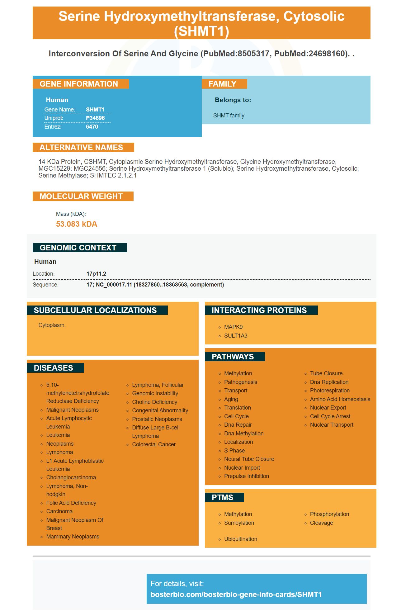


Facts about Serine hydroxymethyltransferase, cytosolic.

| Human | |
|---|---|
| Gene Name: | SHMT1 |
| Uniprot: | P34896 |
| Entrez: | 6470 |

| Belongs to: |
|---|
| SHMT family |

14 kDa protein; CSHMT; cytoplasmic serine hydroxymethyltransferase; Glycine hydroxymethyltransferase; MGC15229; MGC24556; serine hydroxymethyltransferase 1 (soluble); serine hydroxymethyltransferase, cytosolic; Serine methylase; SHMTEC 2.1.2.1
Mass (kDA):
53.083 kDA

| Human | |
|---|---|
| Location: | 17p11.2 |
| Sequence: | 17; NC_000017.11 (18327860..18363563, complement) |
Cytoplasm.


The Boster Bio Anti-SHMT1 marker (catalog number: A02944) is a good choice if you are looking for a new antibody to use in your research. This antibody reacts with all three versions of the SHMT1 protein. The following article will provide more information about clinical applications. We will now discuss the best uses and benefits of the SHMT1 indicator.
Bostern Bio has developed the Anti-SHMT1 marker. It is a high affinity antibody that reacts well with Rat, Mouse, Human. It can be used in ELISA, flow cytometry, and immunofluorescence. It is an integral part of many studies that study human diseases. The instructions for using this antibody with Boster Bio can help you determine if it will react with your research samples.
Hypermethylated repressed gene have been identified by the PANTHER classification. Twenty-six genes from these are involved cell growth, proliferation, and apoptosis. Twenty-six other genes are involved in regulating cellular processes. Six genes concern retinol metabolism. Boster Bio's AntiSHMT1 Marker is responsible for controlling methyl group formation and other transfer reactions.
Data from MeDIP-chip analysis were validated using direct bisulfite sequencing. This validation validated the methylation changes in HCC tissue compared to cancer-free cells. RealTime RTPCR data validated the array-based expression results. These results included seven repressed genes as well as four induced genes. A variety of methods were used to characterize the validated genes, including direct bisulfite and methylation assays.
The recent discovery of CNOT2/9 and SHMT1 in TET patients has shown their prognosis-indicating potential. They can be used as a subtyping marker or alone. Further research will be done to further characterize the proteins and determine if they are potential biomarkers for tumor progression. They will be examined for their potential roles in nucleic acids metabolism and clinical applications.
In vitro studies show that SHMT1 is involved in cell metastasis. HCCLM3 cells expressing SHMT1 showed a higher lung metastasis rate. In nude mice, SHMT1 knockdown significantly increased lung metastasis ability of Hep3B cells. The number of metastatic nodules was significantly higher and they were confirmed by IHC staining as HCC cells.
Knockdown of SHMT1 reduces NOX1 expression, which promotes cell motility and EMT. Similarly, knockdown of NOX1 inhibits the ROS production induced by SHMT1. In the opposite direction, NOX1 restoration reverses ROS-induced cell death. SHMT1 is a promising candidate for biomarkers in HCC, and other types.
The SHMT1 gene is a mitochondrial protein that is involved with the synthesis and maintenance of cellular constituents. It maps to the critical interval in SMS and covers about 40 kb. Twenty-six of 26 SMS patients harbor a deletion of the SHMT1 gene. The 50% SHMT enzyme activity observed in lymphoblasts of patient lymphoblasts is indicative that there is haploinsufficiency. This gene deletion may be responsible for the SMS phenotype, and could point to possible therapeutic interventions.
Many studies have been performed using SHMT1 as a biomarker for clinical use. RNA extracted from RMS cells was used for qPCR with PowerUp SYBR Green Master Mix (Takara Bio) with a Viia7 Real-Time PCR system. The 2-DDCt method was used to determine the relative copies of SHMT1, CDK4 or SHMT2, and the results were compared to their respective values in R cells.
Also, 13 FP RMS PDX cancers showed an increase in the expression of SHMT2 mRNA/protein. The expression of SHMT2 could be normalized to D12Z3 centromeric D12Z3. We measured the SHMT2 concentrations within PDX tumors and in PAX3FOXO1 controls tumors using immunoblotting. ImageJ was used to calculate SHMT2's and GAPDH's protein levels in addition to quantitative analysis. The distribution plot shows how SHMT1 and GAPDH are related in the two groups. To determine the significance of these two groups, the data were statistically examined using Student’s t test in Prism8.
The SHMT1 gene is essential to the folate cycle, but it has not yet been fully inhibited. Boster Bio offers a line of highly targeted antibodies to block this gene. These antibodies have been validated in multiple assays, including Western blotting and Immunohistochemistry. Boster Bio has everything you need to know about this gene.
Selective SHMT1 inhibitors target Cys204. Their action is mediated by 3BP's structure and reactivity. The 3BP enzyme complex inhibits SHMT2's expression. Cys204 was found to negatively regulate SHMT2's activity by inhibiting its binding of substrates. This mutation has the added benefit of reducing MS risk. This is an important step towards developing a drug for Cys204.
A decreased expression of SLC19A1 in cancer cells inhibits folate retention, leading to increased reliance on SHMT1 mediated cytosolic folate flux. Low SLC19A1 levels also reduce cancer cells' ability of metabolizing folate. Patients with low SLC19A1 may have a treatment option by inhibiting SHMT1.
One-carbon metabolism is a hallmark of cancer, and its inhibitors work by restraining tumor formation and progression. It contributes to many biosynthetic pathways, and involves a complex network of enzymes. The two pioneering drugs targeting one-carbon metabolism are methotrexate and 5-fluorouracil. This therapy is still in its infancy and will need to be validated and tested further, despite their success.
Cytoplasmic SHMT1 expression is high in lung cancer. In order to repair DNA replication, tumor cells require cytosolic pathway flux 1C. Thus, tumor cells can grow in nutrient poor environments. Similarly, brain tumor cells have a metabolic switch under poor environmental conditions. They switch to glycolytic phenotype and upregulate the activity of glucose transporters.
PMID: 8505317 by Garrow T.A., et al. Cloning of human cDNAs encoding mitochondrial and cytosolic serine hydroxymethyltransferases and chromosomal localization.
PMID: 9056951 by Chave K.J., et al. Isolation and characterisation of human genomic sequences encoding cytosolic serine hydroxymethyltransferase.


