This website uses cookies to ensure you get the best experience on our website.
- Table of Contents
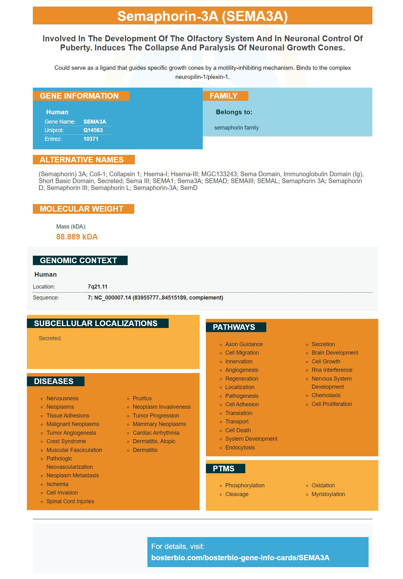
1 Citations 4 Q&As

1 Citations

Facts about Semaphorin-3A.

Could serve as a ligand that guides specific growth cones by a motility-inhibiting mechanism. Binds to the complex neuropilin-1/plexin-1.
| Human | |
|---|---|
| Gene Name: | SEMA3A |
| Uniprot: | Q14563 |
| Entrez: | 10371 |

| Belongs to: |
|---|
| semaphorin family |

(semaphorin) 3A; coll-1; collapsin 1; Hsema-I; Hsema-III; MGC133243; sema domain, immunoglobulin domain (Ig), short basic domain, secreted; Sema III; SEMA1; Sema3A; SEMAD; SEMAIII; SEMAL; Semaphorin 3A; semaphorin D; Semaphorin III; semaphorin L; semaphorin-3A; SemD
Mass (kDA):
88.889 kDA

| Human | |
|---|---|
| Location: | 7q21.11 |
| Sequence: | 7; NC_000007.14 (83955777..84515189, complement) |
Secreted.


PMID: 8269517 by Kolodkin A.L., et al. The semaphorin genes encode a family of transmembrane and secreted growth cone guidance molecules.
PMID: 22416012 by Young J., et al. SEMA3A deletion in a family with Kallmann syndrome validates the role of semaphorin 3A in human puberty and olfactory system development.
*More publications can be found for each product on its corresponding product page


