This website uses cookies to ensure you get the best experience on our website.
- Table of Contents
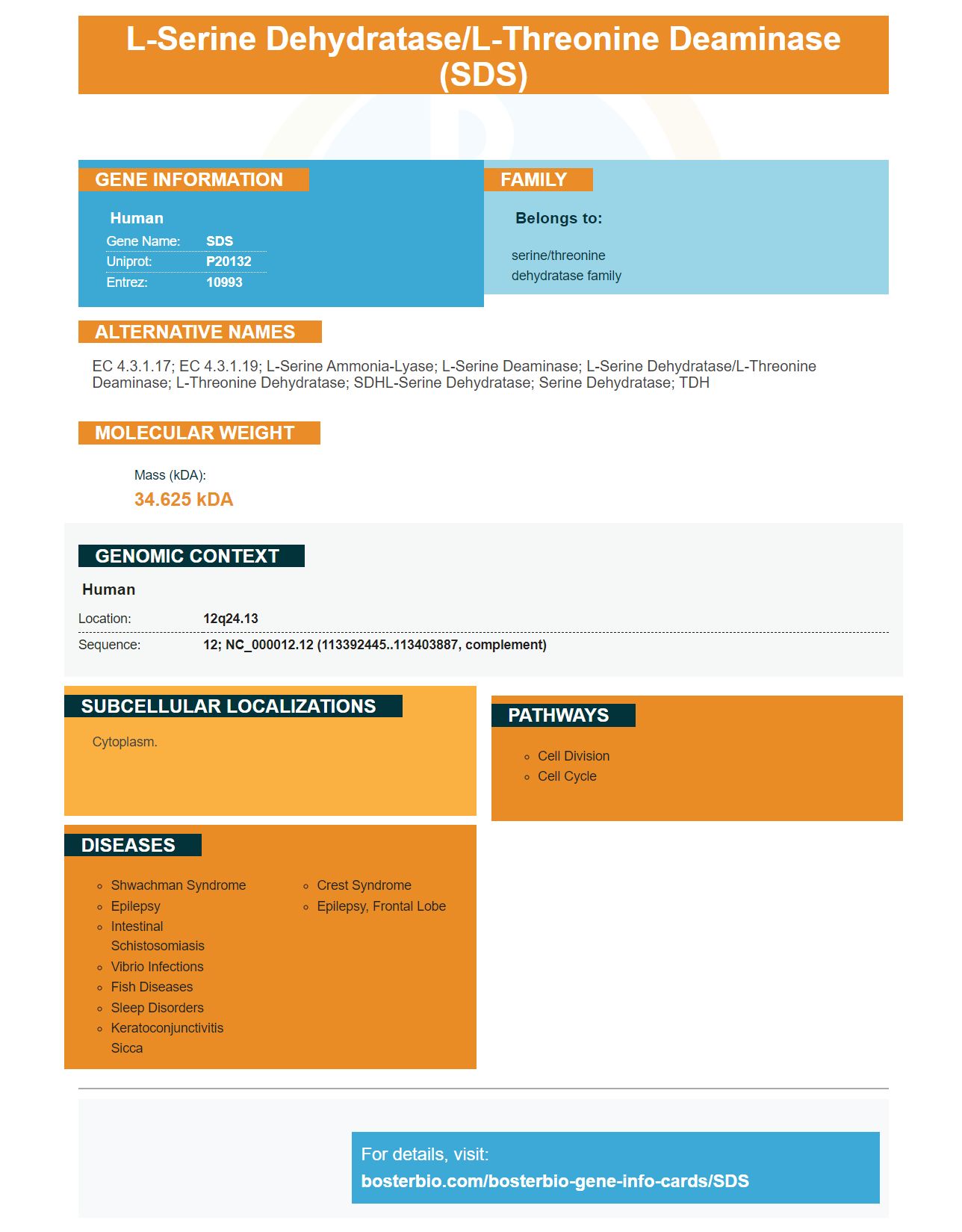
Facts about L-serine dehydratase/L-threonine deaminase.

| Human | |
|---|---|
| Gene Name: | SDS |
| Uniprot: | P20132 |
| Entrez: | 10993 |

| Belongs to: |
|---|
| serine/threonine dehydratase family |

EC 4.3.1.17; EC 4.3.1.19; L-serine ammonia-lyase; L-serine deaminase; L-serine dehydratase/L-threonine deaminase; L-threonine dehydratase; SDHL-serine dehydratase; serine dehydratase; TDH
Mass (kDA):
34.625 kDA

| Human | |
|---|---|
| Location: | 12q24.13 |
| Sequence: | 12; NC_000012.12 (113392445..113403887, complement) |
Cytoplasm.


Boster Bio is one of the brands you've heard of if searching for a protein de-siccator. If not, you might be interested in examining the Dual Color Protein Loading Buffer Super Vision Detection, or High-affinity primary antibodies. These innovative products can help you identify the right protein desiccator for your needs. They can be purchased via Boster Bio's customer support representatives.
As you are aware, the SDS marker is a widely used chemical to detect soluble proteins. However, there are several crucial aspects to consider when preparing your sample for ELISA. First, you need to prepare the sample in the best possible manner. Concentrate the antibodies in a lysis buffer to decrease the degree of cross-linking. Additionally, you must select the best blocking buffer. Boster Bio also offers an optimized ELISA guide to aid you in obtaining the best results from your study.
The SDS marker is essential for immunohistochemistry, which uses the principle of antibodies binding to antigens within a sample. The guide also provides advice on how to conduct optimal tests using immunohistochemistry. It also provides details on sample preparation and troubleshooting. There are a variety of technical sources blogs, articles, and information on diseases on Boster's website. The SDS marker is a vital element of many immunostaining procedures.
The Dual Color Protein Loading Buffer includes Bromophenol blue to prevent the degradation of proteins during heating. This buffer is particularly beneficial for SDS-PAGE as many proteins are sensitive to pH changes caused by the temperature changes in Tris buffers. It also contains two tracking dyes: Blue (Bromophenol blue) and Pink ("Pyronin Y") to monitor the transfer of proteins to the membrane. The buffers can be stored at -20°C for several months and four degrees Celsius before use.
For the detection of proteins of interest, researchers have developed the primary-secondary-ABC system. This system employs avidins as well as the signal molecule, which is typically Horseradish Peroxide, HRP. Researchers can find proteins of interest that have high specificity, low background and high titration based on the resulting Titration. Other detection systems are based upon polysaccharides and organic polymers. Boster Bio's Super Vision Detection kits are among these advanced systems.
The Dual Color Protein Loading buffer is a complete buffer system that helps reduce temperature changes and pH fluctuations during SDS/PAGE analysis. This buffer solution contains two tracking dyes such as pyroninin Y, and bromophenol blue, in conjunction with SDS and DTT. The buffer is formulated to provide the optimal amount of proteins as well as nutrients required for success in Western Blot analysis.
The buffer is designed to reduce SDS and allow loading of low molecular weight proteins. The RIPA lysis buffer may also be used for purification of proteins or western blotting. BosteraEUR(tm), IP Lysis Buffer, is a mammalian whole-cell lysis buffer that does not contain SDS. It is based on a modified RIPA buffer formula. The WST-1 Cell Proliferation Assay Kit allows users to assess the viability and proliferation of cells in a culture.
Boster Bio is a world leader in the development and manufacture of ELISA kits, research antibodies, and is dedicated to supporting science through using reagents of the best quality. Their products are optimized for efficiency and picogram-level sensitivity, making it possible for researchers to detect biomarkers in a variety areas such as neurosciences, cancer, inflammation, and developmental biology. In addition to their line of ELISA kits, Boster offers a range of high-affinity primary antibodies and ELISA kits for research.
In the context of immunoglobulins, primary antibodies are composed of specific antibodies that only bind to the antigen being targeted. Their quality is determined by two variables such as affinity and specificity. A high affinity antibody is able to bind strongly to the targeted antigen and a lower specificity means it may be able to bind to non-targeted antigens. This ensures that antibodies are able to detection the purification process, as well as the measurement of their targets.
For instance, Boster's antibeta actin rabbit monoclonal antibody has been used as an internal control for western blotting, to examine the expression of retinol binding proteins 4 in the granulosa cells of porcine. Boster's high affinity secondary antibodies can be used to purify specific types of antibodies. Boster produces an array of special antibodies in addition to primary antibodies.
A wide range of secondary antibodies can be used in ELISA to identify the same antigen. Dual antibodies reduce cross-reactivity, and require an additional incubation process. This technique is highly sensitive, however it requires further optimization in order to reduce cross-reactivity. No matter which antibody is used to test the antigen's specificity an assay with two assays will prove the most efficient. This permits researchers to study various matrices, including cells, virological or tissue samples.
Boster Bio SDS Marker Troubleshooting Guide offers tips and procedures to optimize flow cytometry experiments. The first step to achieving the best results is sample preparation. This troubleshooting guide gives insight into sample preparation including antigen retrieval and fixation embedding as well and other subjects. Below are some of the most common sample preparation mistakes and tips to fix them.
PMID: 2674117 by Ogawa H., et al. Human liver serine dehydratase. cDNA cloning and sequence homology with hydroxyamino acid dehydratases from other sources.
PMID: 18342636 by Yamada T., et al. A catalytic mechanism that explains a low catalytic activity of serine dehydratase like-1 from human cancer cells: crystal structure and site-directed mutagenesis studies.


