This website uses cookies to ensure you get the best experience on our website.
- Table of Contents
2 Citations 7 Q&As
Facts about Syndecan-3.
.
Human | |
---|---|
Gene Name: | SDC3 |
Uniprot: | O75056 |
Entrez: | 9672 |
Belongs to: |
---|
syndecan proteoglycan family |
KIAA0468; N-Syndecan; SDC3; SDCN; SYND3syndecan proteoglycan 3; syndecan 3 (N-syndecan); syndecan 3; syndecan neural type; Syndecan3; Syndecan-3
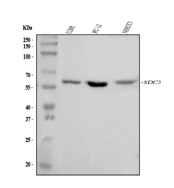
Mass (kDA):
45.497 kDA
Human | |
---|---|
Location: | 1p35.2 |
Sequence: | 1; NC_000001.11 (30869466..30909735, complement) |
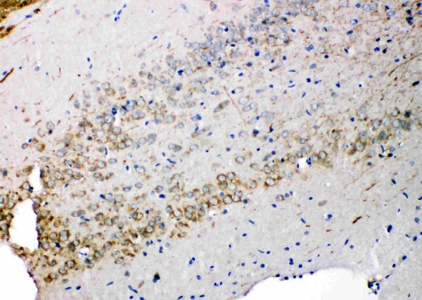
Expressed in the nervous system, the adrenal gland, and the spleen.
Cell membrane; Single-pass type I membrane protein.
There are many questions to be asked about optimizing experiments using Boster Bio. Boster Bio optimization guides will provide the complete information you require to optimize your experiments and improve outcomes. However every experiment is not perfect. Even though most researchers have issues with their research due to inadequate controls, they can be eliminated. Troubleshooting guides can assist you to solve these issues. These guides are extremely helpful for all researchers, regardless of whether they're working in biology or not.
SDC3 is a protein that is present in inflamed vascular endothelia. It has numerous functions, including interacting with leukocytes and inflammatory chemicals. These tissues are high in levels of SDC3, which can suggest a cause-and-effect relationship. SDC3 is being used by researchers to diagnose diseases of inflammation and for research.
SDC3 is also a crucial factor in inflammatory reactions. Cells produce SDC3 when TNF-a is added. To determine the levels of the protein, imaging flow cytometry was employed. This study was comprised of ten independent studies. The results suggest that SDC3 is involved in inflammatory diseases such as Alzheimer's. The role of SDC3 in inflammation is still unknown.
SDC3 is a new chemical that is found in various human tissues, including in the central nervous system and brain. SDCs regulate inflammation and interact with leukocytes in order to facilitate exovascularization. SDC3 is found in peripheral blood monocytes as well as primary brain endothelia. The high level of expression is associated with amyloid plaque accumulations in the brain. This suggests that SDC3 could be a biomarker that could aid in early detection and treatment of Alzheimer's disease.
The SDC3 immunostaining antibody detects Iba-1 (green) and CD45 (red) microglia within the brain. Contrary to CD45-positive cell types and clusters of CD45-positive microglia Iba-1-positive microglia cluster around amyloid deposits. They have thinner, smaller processes, and higher Iba-1 levels than microglia which are not present.
The results of this study indicate that a tiny subset of SDC3-positive cells are able to differentiate between microglia and neuronal cells, and that their staining differs in areas that have amyloid plaques. Iba-1a positive cells were more frequent near amyloid plaques. However CD45-positive cells showed higher immunoreactivity in the brain parenchyma, blood vessels, and meninges.
The study was published in the FASEB Journal by Abramowski DC. Both the soluble Ab40 and insoluble Ab40 levels were both elevated. The insoluble Ab42 level was not statistically significant. Additionally, serum Ab40 levels did not differ between the two groups. It has also been proven that SDC3 immunostaining of amyloid plaques does not affect cognitive functions.
The relationship between TNF-a levels and the presence of NS is highest in the northern hemisphere. However, the sensitiveness of this biomarker to determine NS is less sensitive in the south hemisphere. This is why it should be treated with caution. The study found that TNF-a has moderate diagnostic sensibility and a high specificity when compared to PCT and CRP. Its positive correlation with NS is in line with its use as a biomarker for diagnosis of NS.
In Indonesia this study was conducted on an insignificant population. The levels of TNF-a and sVCAM-1 levels did not correlate. A causal relationship between the variables was also not established. In light of this, it isn't feasible to draw any definitive conclusions regarding the role played by TNF-a in RA. The study should be re-examined with larger numbers of patients and controls. These findings are nevertheless encouraging.
The serum levels of TNF-a are negatively associated with the eGFR. However there isn't a significant relationship between TNF levels and the serum IgA and the count of red blood cells in the urine. However TNF-a levels appear to be positively correlated with 24-h urinary protein excretion and ratio of urinary protein to serum creatinine. Moreover, serum TNF-a is positively associated with urinary protein excretion and Cystatin C levels.
TNF-a is involved in a variety roles. It is involved in numerous pathological and homeostatic processes. It is involved in a variety of functions and, in addition to being a powerful paracrine molecule, TNF-a plays an important role in controlling the activity of various cell types. Additionally, it aids in the development of insulin resistance and dyslipidemia. These factors have been found to play a major role in inflammatory conditions, including atherosclerosis.
TNF-a is structurally linked to lymphotoxin. Both are active when the membrane is intact, but they carry out different functions. They must be studied to determine their respective roles. The relationship between lymphotoxin and TNF-a isn't yet conclusive. The question is "Is TNF a cytokine related?"
TNF-a is well-known for its pro-inflammatory effects, however, C3 is also associated to Apoptosis or the emergence of the apoptosis. TNF-a regulates blood coagulation and plays a key role in the disruption or circulation. It also increases the amount of oxidative stress. The correlation between TNF-a and apoptosis is related to altered coagulation propensity.
The levels in the blood of TNF-a as well as the IL-6 are greater in those suffering from hypertensive renal nephropathy than those who suffer from normoalbuminuria or proteinuria. Lower serum levels are associated to a higher incidence of NS. Both proteins could play a crucial role in the development and pathogenesis of NS. As we continue to gain knowledge about the NS further studies are needed to determine if the two proteins are somehow linked.
There is also a connection between the maternal weight and the levels of leptin in plasma. It is not significant for both the first however it does tend to increase in the former. There was no correlation between TNF-a levels and fetal leptin concentrations. This suggests that the placenta could be the main driver in the transport of leptin from mother to child. But, maternal adiposity did not affect the levels of TNF and IL-1b in the fetus or the mother. This could be due to the fact that the cytokines are localized and controlled.
PMID: 11527150 by Berndt C., et al. Cloning and characterization of human syndecan-3.
PMID: 9388509 by Asundi V.K., et al. Phosphorylation of recombinant N-syndecan (syndecan 3) core protein.
*More publications can be found for each product on its corresponding product page