This website uses cookies to ensure you get the best experience on our website.
- Table of Contents
1 Citations 8 Q&As
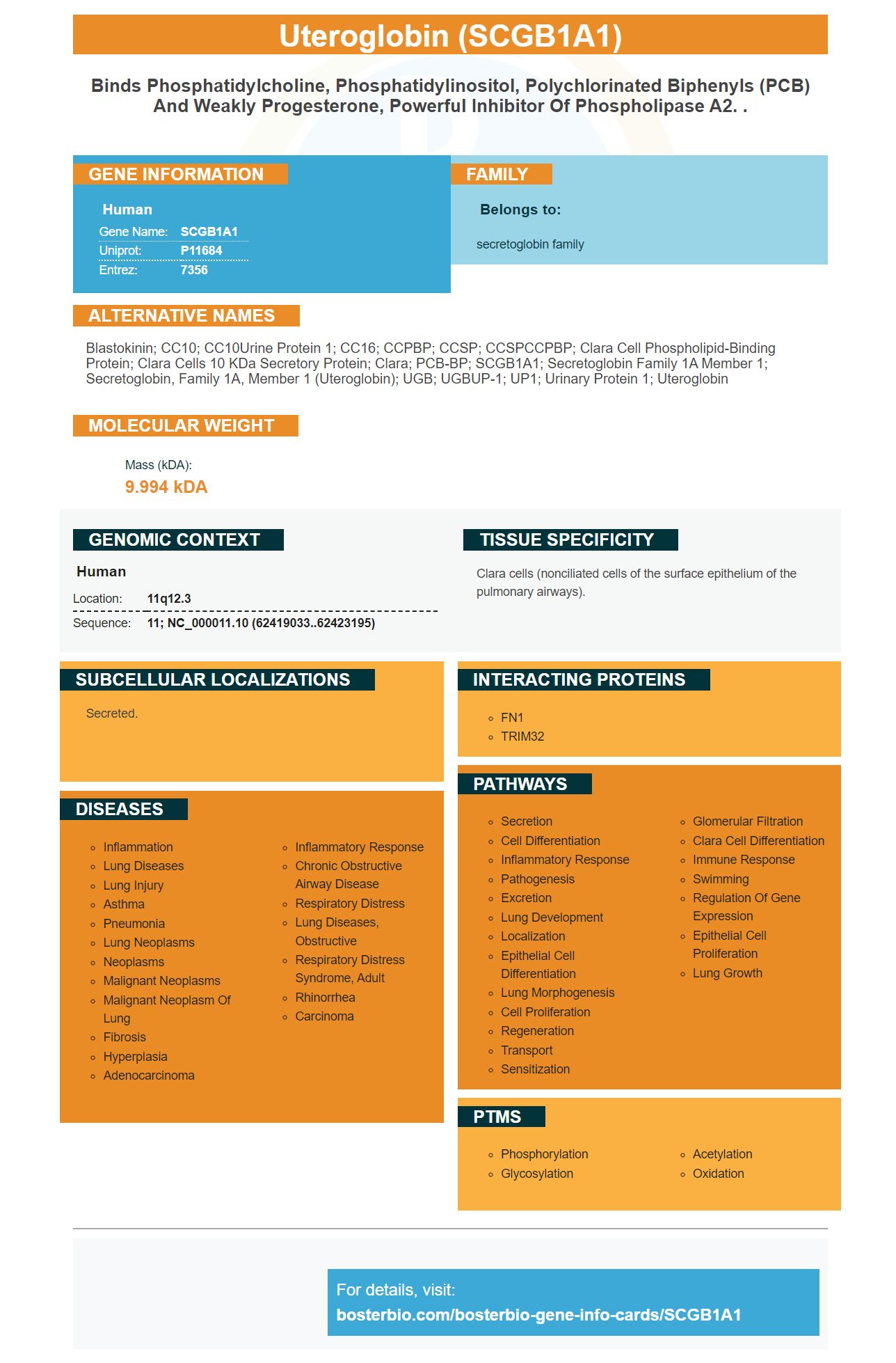
Facts about Uteroglobin.
Human | |
---|---|
Gene Name: | SCGB1A1 |
Uniprot: | P11684 |
Entrez: | 7356 |
Belongs to: |
---|
secretoglobin family |
Blastokinin; CC10; CC10Urine protein 1; CC16; CCPBP; CCSP; CCSPCCPBP; Clara cell phospholipid-binding protein; Clara cells 10 kDa secretory protein; Clara; PCB-BP; SCGB1A1; Secretoglobin family 1A member 1; secretoglobin, family 1A, member 1 (uteroglobin); UGB; UGBUP-1; UP1; Urinary protein 1; Uteroglobin
Mass (kDA):
9.994 kDA
Human | |
---|---|
Location: | 11q12.3 |
Sequence: | 11; NC_000011.10 (62419033..62423195) |
Clara cells (nonciliated cells of the surface epithelium of the pulmonary airways).
Secreted.
This article will describe how to prepare a Cytospin preparation that includes the SCGB1A1 marker, as well as the immunoostaining of EGFP+ cells within the tracheal epithelium and other uses for the SCGB1A1 reagent. The information presented is applicable to every scientist around the globe. Below are some examples of beneficial experiments that have been performed with the SCGB1A1 marker.
The present study utilized an SCGB1A1 cytospin preparation to determine naive cell types. The number of cells per mouse was determined and differential cell counts were performed using Wescor cytospin preparations stained with DiffQuick. The cell trajectory of BEGM was different from those of MCCs. In both genotypes, a greater percentage of SCGB1A1+ cells was observed.
The presence of transitional cells was observed in the trachea of newborn animals and in the homeostatic bronchi of human beings. This marker is immunodetectable in human bronchi. SCGB1A1-positive cells were found in the human trachea. Cytospins prepared from SCGB1A1positive cells were made by using the lectin UEA-1, which is an anti-muc5ac rabbit polyclonal Muc5ac, and lectin UEA-1.
The most effective marker gene for club and goblet cells was identified using scRNA-seq and ALI 28 differentiation medium. Additionally, the bars on top of the heatmaps reflect the cell type and pseudotime. The cells were split by type and the resulting RNA analysis revealed the types of cells. The results also showed that cell dissociation has no effect on gene expression.
The authors employed a green fluorescent proteins (GFP) mouse strain to increase EGFP expression in nonciliated and ciliated cells in mice with EGFP-positive genes. The fluorescent protein is produced only by ciliated cells in trachea. It is not expressed by other kinds of cells or tissues within the lung. In the trachea however GFP-positive cells were observed specifically in the respiratory epithelium, which is located in the paranasal sinuses as well as the nose. These cells are situated in the transition region between the respiratory epithelium as well as the olfactory epithelium, making them easily visible.
This immunostaining demonstrated that the majority of the epithelium of the olfactory sense was negative. Some cells were positively stained, such as small round cells with a tube-like extension, similar to neuroepithelial cells that regenerate. The immunostaining of EGFP+ cells in the tracheal epithelium was performed with the BD FACSAria II cell sorter. Sorting gates were positioned on the low-SSC/eGFP+ cells. After sorting the cells, samples were placed in HBSS buffer until they were processed using RT-PCR experiments. Data acquisition and analysis was done using BD FACSDiva Software by BD Biosciences PharMingen.
The resultant immunostaining for EGFP+ cells in the tracheal tissue was conducted on ChAT-eGFP-expressing mice. In the same way, ChAT-eGFP-positive cell lines were isolated from freshly dissociated epithelial tissue. After removal, RT-PCR analysis was conducted to confirm the presence of ChAT-eGFP+ cells. ChAT-eGFP also validated expression of other genes, for instance, PGP 9.5 or the heavy chain myosin.
ChAT-eGFP+ cells showed ultrastructural eGFP immunohistochemistry. They also showed the immunoreactivity of VAChT, a membrane protein that binds cholinergic Vesicles. VAChT-IR was identified in a subset ChAT-eGFP+- cells in the tracheas of wild type. However, the amount of immunoreactivity found in VAChT positive cells was significantly enhanced after eGFP immunolabeling.
The Boster Bio Anti-p62 Protein (BBI-AP62) was created to focus on the nuclear import of a molecule that is capable of interfacing with c-Jun. The protein binds to the SH2 domain of the lck. It also has two possible NLSs on amino acids 186-191 and 264-270. These residues can be altered to inhibit nuclear import 1.5-fold or 6.3-fold according to their mutation.
Autophagy is a crucial process in our cells, and the ubiquitinated cargo p62/SQSTM1 binds to be degraded by Lysosomes. Autophagy is regulated by MAP LC3A, but its level does not reflect the autophagic flux. Because this protein is part of the autophagic pathway, it can be useful in analyzing the flow of the process.
Boster Bio Antip62 Protein (BPIAP) targets Sequestosome-1. This gene is encoded by SQSTM1. It is also known as p60, a170 or dynactin subunit 4. The antibody targets both unrelated and related proteins. The website of the manufacturer lists the target immunogen and the applications that have been tested.
The mCherry P62 tagged with a mCherry is a useful tool for studying nuclear import. The NLS2 protein in the mCherry-tagged protein controls nuclear import. The mCherry-tagged mutant p62-K7A/D69A mutated Threonine 269 increased nuclear accumulation by 1.7 times. It also inhibited the p62 import, which blocked nuclear import.
The SQSTM1 marker is useful in various fields. It is an important marker in cells, particles and tissue samples. Boster's anti-p62 Protein (SQSTM1) antibody is a highly sensitive marker that is widely utilized in a variety of research projects. The company is constantly improving this kit to make it as adaptable as possible. It has been proven to be an effective instrument for researchers and biotech companies too.
Desmoglein-1, a transmembrane protein found in the desmosome, is also known as Desmoglein-1. It is part of the cadherin family. It is believed to play a role in cell-cell adhesion. Desmoglein is a blend of the Greek words "desmos", "glein" and "glue-like". The molecule is found in all tissues that have desmosomes. It is linked to intermediate filaments in cytoskeleton by a variety of calcium binding sites. Desmogleins play an important function in maintaining cell stability and are involved in the progression of cancer.
The anti-Desmoglein 2+2 antigen was detected in blood donors from Tunisia as well as in patients suffering from pemphigus foetus. Both groups' serum samples were subjected to immunoassay with antigen-coated microtitre plates. The samples were then washed with goat antimouse IgG. Twenty-five positive ELISAs were processed to determine the presence of desmoglein antibodies. In addition an indirect immunofluorescence substrate was utilized.
In a model pemphigus based on baculoproteins foetus, the antigen that corresponds to the extracellular domain Dsg1 was discovered. The antigen has been reported in various publications and identified as an autoantigen via the study of immunochemicals. The study also revealed that DSG1 plays a significant role in adhesion of cells.
DSG1 was once thought to be a structural adhesion molecule. However, recent research has demonstrated that DSG1 expression is more important than cell adhesion. Furthermore, misexpression of DSG3 causes an abnormal stratum corneum permitting more transepidermal aqueous to leak. DSG2 is also involved in the formation and spread of tumors within the suprabasilar layer.
This monoclonal antibody identifies peptide the peptides 181-195 of synaptopodin in the mouse brain. This is in line with amino acids 188 - 202 in synaptopodins of humans. The antiserum was derived by immunizing rabbits with keyhole limpet hemocyanin-conjugated peptides. Utilizing an affinity-purified peptide the antiserum was purified with Ultralink technology which is a commercially-available protein purification technology.
Synaptopodin is an important protein expressed in a subset of neurons in the brain. The solubility of this protein was examined by conducting fractionation experiments using two buffers which are low salt and high salt and a mAb to detect synaptopodin. The mAb binds synaptopodin to around 80percent.
This antibody is able to detect synaptopodin through immunofluorescence. Eight-mm-thick frozen sections of perfusion fixed tissue were used to study synaptopodin's distribution. The protein is present in the cerebral cortex, the striatum, hippocampus, and Olfactory bulb, however no other regions were identified. Anti-Synaptopodin monoclonal antibody against the SCGB1A1 marker was designed to recognize synapses.
Synaptopodin gene expression is differentiation-dependent. It is first apparent in the rat brain at the 15th day of postnatal maturation and increases until it reaches its peak at the age of adulthood. In-situ hybridization confirmed the protein synthesis, and it was found to be restricted to the cerebral cortex and the hippocampus. It is possible that synaptopodin is playing significant functions in regulating dendritic spinal shape and podocyte foot processes.
Synaptopodin is linked to actin. Double labeling experiments using the antibody against actin revealed synaptopodin to be closely connected to this structural component. It was found to interact with cytochalasin B and F-actin. Depolymerization of microtubular network did not significantly affect the distribution of synaptopodin, nor did it alter the pattern.
PMID: 3167058 by Singh G., et al. Amino-acid and cDNA nucleotide sequences of human Clara cell 10 kDa protein.
PMID: 7733299 by Hay J.G., et al. Human CC10 gene expression in airway epithelium and subchromosomal locus suggest linkage to airway disease.
*More publications can be found for each product on its corresponding product page