This website uses cookies to ensure you get the best experience on our website.
- Table of Contents
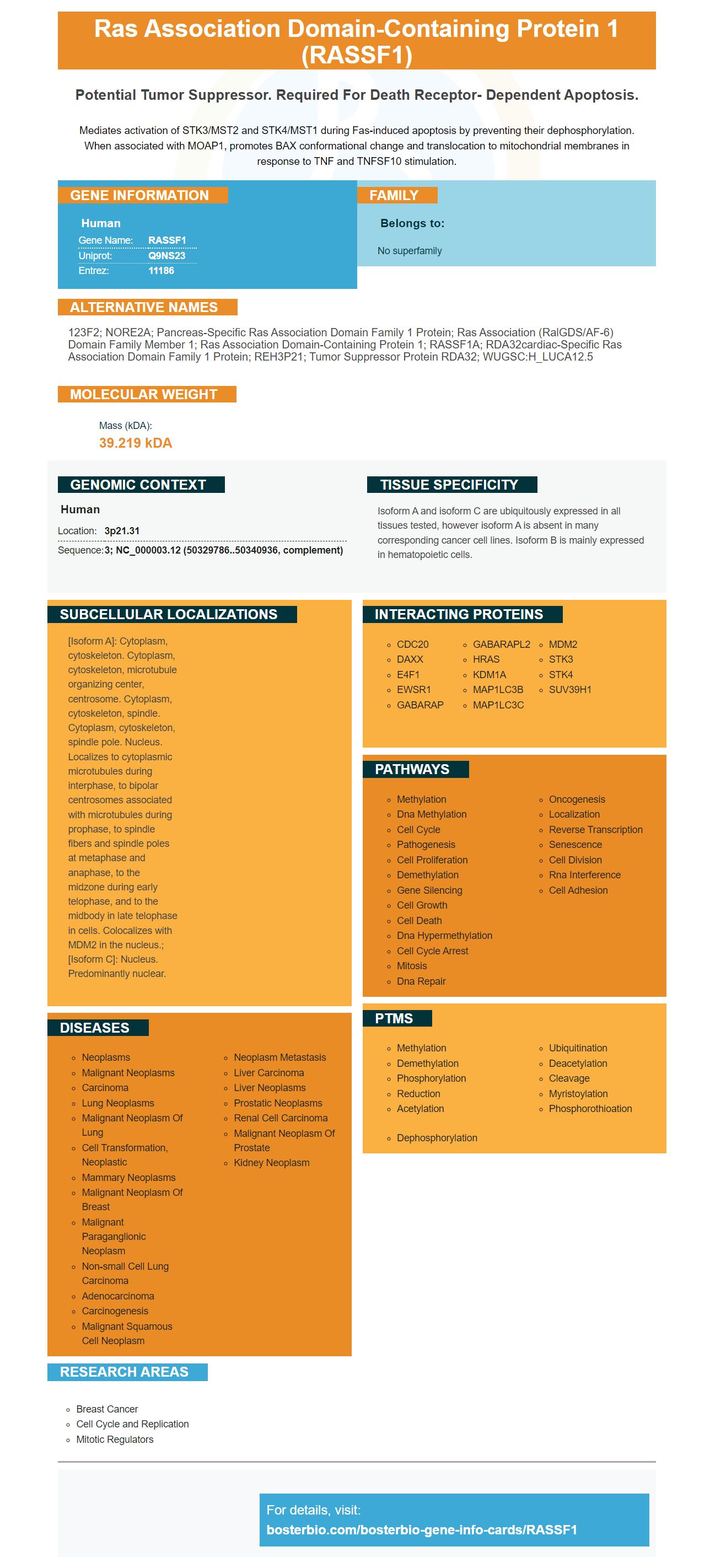
Facts about Ras association domain-containing protein 1.
Mediates activation of STK3/MST2 and STK4/MST1 during Fas-induced apoptosis by preventing their dephosphorylation. When associated with MOAP1, promotes BAX conformational change and translocation to mitochondrial membranes in response to TNF and TNFSF10 stimulation.
Human | |
---|---|
Gene Name: | RASSF1 |
Uniprot: | Q9NS23 |
Entrez: | 11186 |
Belongs to: |
---|
No superfamily |
123F2; NORE2A; pancreas-specific ras association domain family 1 protein; Ras association (RalGDS/AF-6) domain family member 1; ras association domain-containing protein 1; RASSF1A; RDA32cardiac-specific ras association domain family 1 protein; REH3P21; tumor suppressor protein RDA32; WUGSC:H_LUCA12.5
Mass (kDA):
39.219 kDA
Human | |
---|---|
Location: | 3p21.31 |
Sequence: | 3; NC_000003.12 (50329786..50340936, complement) |
Isoform A and isoform C are ubiquitously expressed in all tissues tested, however isoform A is absent in many corresponding cancer cell lines. Isoform B is mainly expressed in hematopoietic cells.
[Isoform A]: Cytoplasm, cytoskeleton. Cytoplasm, cytoskeleton, microtubule organizing center, centrosome. Cytoplasm, cytoskeleton, spindle. Cytoplasm, cytoskeleton, spindle pole. Nucleus. Localizes to cytoplasmic microtubules during interphase, to bipolar centrosomes associated with microtubules during prophase, to spindle fibers and spindle poles at metaphase and anaphase, to the midzone during early telophase, and to the midbody in late telophase in cells. Colocalizes with MDM2 in the nucleus.; [Isoform C]: Nucleus. Predominantly nuclear.
Boster Bio Anti–RASSF1 Monoclonal Antibody reacts against Human. It is stable at -20degC (for up to one-year) or at 4degC (for one month). It is derived using purified human RASSF1-expressing E. coli protein. The antibody can be blocked by purchasing a blocking peptide, which varies in cost based on the length of the immunogen. The Boster Bio Anti-RASSF1 Monoclonal Antibody is tested on known positive and negative samples.
RASSF1 was detected in rat mammal cancer by IHC staining. This antibody binds the RASSF1 protein. It is expressed in cells of the mammary ductal lobe. In this study, we used three-week-old rats with tumors. Inoculation with four neoplastic line cells. In addition, we used syngeneic F344 rats to perform these experiments.
We have used a rat mammary carcinoma model to study the role of this RNA in breast cancer. It was previously reported that the RASSF1 gene plays a crucial role in regulating tumor growth and metastasis. It has been shown that it plays a key role in regulating HER2 levels in breast cancer. This mRNA has been successfully used by researchers to perform IHC analysis.
The RASSF1A protein plays a significant role in the physical properties and progression of cancer-associated ECM. Additionally, the loss of RASSF1A may cause disruptions in collagen organization. This loss in RASSF1A was studied by analysing the structural integrity h2299control cells embedded into non-crosslinked collagen matrix.
In a separate experiment, we used human lung adenocarcinoma cells (h2299TetON-pcDNA3-RASSF1A) as a model for rat mammary carcinoma. RASSF1A gene expression was stimulated with l-glutamine and doxycycline.
It is interesting to note that both T. vulgaris (and the RASSF1 marker) can effectively suppress the development mammary tumors in a rodent model. The chemoprevention T. vulgaris prevented the mammary gland from growing, but it did not affect the incidence or latency of tumors in the rat model. Further, the T. vulgaris-induced rat mammary tumors decreased the size of the tumors by 43.5%, indicating that T. vulgaris is a potent anticancer drug that can have therapeutic effects in the human population.
Recent research has shown IHC staining for colorecTAL can be used to detect tumors with high RASSF1 levels. This marker is expressed within the nucleus in a subset or melanoma-cell cells. Its expression is measured in the core of the cancer and in the buds. This is a valuable tool to evaluate colorectal tumors.
This study examined the relationship between RASSF1-methylation and early regression. It was found that APC, CDh23, p16 and CDh23 methylation were associated with a higher risk of recurrence. The absence of methylation was associated with a 25-month median time to recurrence.
The IHC staining of RASSF1 can be used to classify CRC based on the expression of RASSF1 protein and a variety of other genes. This can help clinicians determine whether a patient has colorectal carcinoma. In addition, maspin expression is helpful for assessing the extent of EMT in CRCs. Nuclear positivity is used to identify the mesenchymal Subtype and may also indicate lymphoma metastasis. Further research is needed in order to determine the clinical significance of RASSF1 protein IHC staining.
PDRG1 is a protein that is overexpressed in colon cancer. Western blotting was performed using protein samples taken from normal as well as tumor tissues. Anti-actin antibody was used for loading and determining RASSF1 levels. This study also suggests that RASSF1 may be an effective marker for colorectal cancer. This study has proven that RASSF1 is a reliable marker for PDRG1 excess.
There are many ways to identify prostate cancer using IHC. The most common method to identify prostate cancer is to check for the PSA markers. However, this may not always be accurate. PSA might not be elevated in some cases. Other methods may include PSMA, HOXB13, or prostein. Before performing the biopsy, it is important to understand what each marker looks like.
Researchers tested 131 cases of prostate cancer, 61 benign samples of prostate hypertrophy, and human prostatic cell lines to determine whether this marker is useful in early detection. Tests for prostate cancer showed lower levels of RASSF1A compared to control tissue. Additionally, there was a significant decrease in tumor proteins in metastatic lesions. These findings were associated to higher Fuhrman's grades and TNM grades.
The RASSF1 marker is not useful in diagnosing prostate cancer. However, it does indicate that keratin 903 or p63 can be used as adjuncts to the diagnostic process. Kristiansen G, and Sailer V developed these antibodies. They are both members of IIiDUPG & Sailer V, respective. The results are consistent in other studies using these marker, but further testing will be needed to determine whether they are superior.
IHC staining prostate cancer using RASSF1 can be helpful in detecting metastatic lesions, particularly in small tumor foci. Using this marker may help doctors to determine whether tumors are small or metastatic. IHC tests may also help determine if the cancer is spreading. They can help diagnose the disease earlier and predict the progression of the disease.
There are several limitations associated with prognostic immunohistochemistry. Although it can provide prognostic information regarding the relevance of proteins to tumors, it has not been capable of integrating itself into the diagnostic workflow. Furthermore, there are issues of reproducibility, which hamper reproducibility of cutoff values. IHC staining is useful for determining the stage of cancer. However, it cannot replace conventional pathology or reference histology. Further research is required to determine if IHC staining can be used to predict the response to therapy.
Thymic Neoplasms of Entropy are uncommon and account for less that 5% of all thymic tumours. These tumors are often carcinoid in nature, and they affect male patients. Thymic high-grade tumors are less common than carcinoid thymomas and are not associated to tobacco smoking.
This type of cancer is characterised by high-grade nuclear morphology and multiple foci for blood vascular invasion. It has a high mitotic rate and is usually accompanied by a high ki67 index. This type of tumor also has a low level of necrosis. Thymic NETs are not the same as other types of cancer. The diagnosis can be based on only histological findings or in combination with other types.
Thymic tumors can be distinguished from other types by IHC staining. However, a large number of thymic tumours do not show signs of malignancy. A positive CK17 antibody does not rule out the possibility for malignancy. However, the authors acknowledge that their study has limitations.
Despite being rare, thymic neuroendocrine tumors are similar in general behavior and genetic characteristics to their pulmonary counterparts. They will likely be classified in the same way as other pulmonary neuroendocrine cancers. However, there are many unanswered questions and need further research in order to understand their immunohistochemical and molecular differences. Because these tumors have few similarities with their pulmonary counterparts, they require a coordinated international effort to identify them correctly.
Only a handful of immunohistochemical markers currently allow for the detection and diagnosis of thymic net. Tissue microarray analysis was done in a single case with thymic net. The results showed that MUC-1, c-kit, and c–kit were particularly effective in distinguishing thymic cancer from other types. The staining scores were calculated based upon the intensity and percentage positive cells.
MEN-1 is a risk factor for thymic NET. MEN-1 accounts for 25% of all thymic NETs. It is interesting to note that there is no age difference between a sporadic thymic NET or a MEN-1-associated cancer. The majority thymic networks are male. However, there are not many published cases of MEN-1 tumors.
PMID: 10888881 by Dammann R., et al. Epigenetic inactivation of a RAS association domain family protein from the lung tumour suppressor locus 3p21.3.
PMID: 11333291 by Burbee D.G., et al. Epigenetic inactivation of RASSF1A in lung and breast cancers and malignant phenotype suppression.