This website uses cookies to ensure you get the best experience on our website.
- Table of Contents
16 Q&As
Facts about Runt-related transcription factor 3.
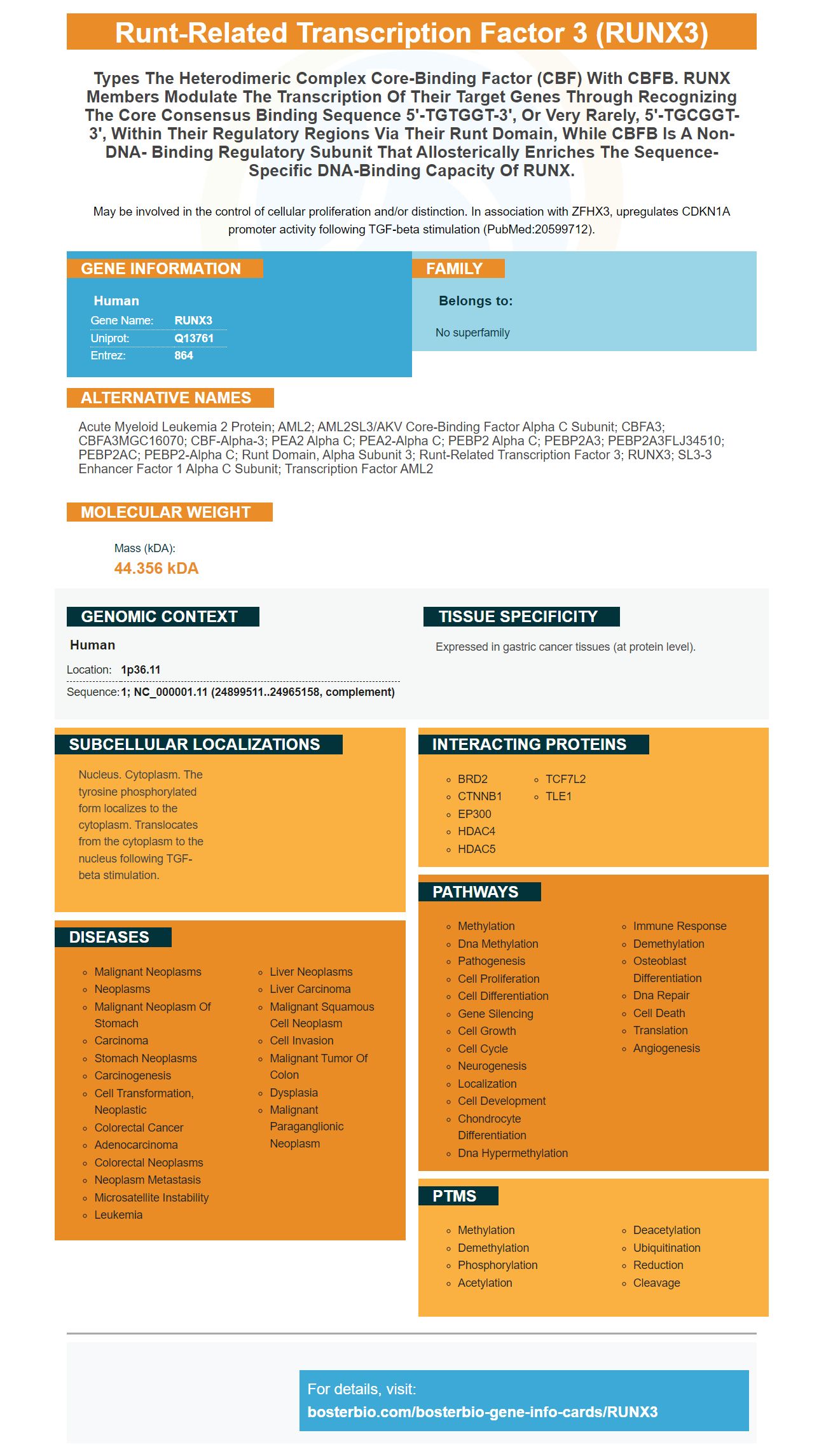
May be involved in the control of cellular proliferation and/or distinction. In association with ZFHX3, upregulates CDKN1A promoter activity following TGF-beta stimulation (PubMed:20599712).
Human | |
---|---|
Gene Name: | RUNX3 |
Uniprot: | Q13761 |
Entrez: | 864 |
Belongs to: |
---|
No superfamily |
Acute myeloid leukemia 2 protein; AML2; AML2SL3/AKV core-binding factor alpha C subunit; CBFA3; CBFA3MGC16070; CBF-alpha-3; PEA2 alpha C; PEA2-alpha C; PEBP2 alpha C; PEBP2A3; PEBP2A3FLJ34510; PEBP2AC; PEBP2-alpha C; runt domain, alpha subunit 3; runt-related transcription factor 3; RUNX3; SL3-3 enhancer factor 1 alpha C subunit; transcription factor AML2
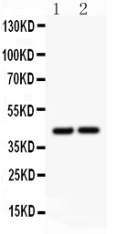
Mass (kDA):
44.356 kDA
Human | |
---|---|
Location: | 1p36.11 |
Sequence: | 1; NC_000001.11 (24899511..24965158, complement) |
Expressed in gastric cancer tissues (at protein level).
Nucleus. Cytoplasm. The tyrosine phosphorylated form localizes to the cytoplasm. Translocates from the cytoplasm to the nucleus following TGF-beta stimulation.
Boster Bio: What are the most beneficial uses of The RUNX3 Protein Protein? RUNX3, which was developed by Steven Boster in 1993, has been linked to invasive bone metastases of oral cancer. The protein is believed to be a valuable biomarker in oral cancer, especially when it is in combination with PTHrP and TGF-b. What is the protein , and how can it help your lab? Learn more in this article.
RUNX3 regulates TRM CD8+ cell differentiation. Runx3 was required for TRM cell populations to be established in a variety of tissue environments, and supported genes involved in tissue exodus and recirculation. Runx3's activity was also associated with the TRM gene expression signature of both mouse and human lymphocytes that invaded tumors. When Runx3 was absent in mice, TRM cells failed to grow in tumors, and the mice had higher rates of tumor growth.
In addition, RUNX3 expression negatively correlated with the LNM and T of ESCC patients. These results were also confirmed by transwell assays in which cells transfected with RUNX3 DNA plasmids displayed decreased invasion and migration. The plasmids slowed TGF-b1-induced EMT from ESCC cell lines, and saved the cisplatin-resistant ESCC cells. Therefore, ESCC is being treated with new medications that target RUNX3
Recent research has revealed that RUNX3 downregulation is associated with early carcinogenesis. Since the first stages of carcinogenic diseases are characterized by downregulation of RUNX3 It has been hypothesised that the suppression of RUNX3 may hinder cell invasion. In addition to that, reducing RUNX3 expression in cancerous cells of the gastric tract could aid in preventing the recurrence of the disease.
The researchers of this study propose that RUNX3 is involved in the dorsal root ganglion's axonal projection. Additionally, RUNX3 regulates the expression of cytoskeletal proteins. It also has been linked to various kinds of cancer. RUNX3 mutations have been associated with gastric cancer in several studies. The authors conclude that the results of these studies suggest that RUNX3 could be beneficial to the treatment of cancers in humans and animals.
Although the antitumor effect is not yet established, exogenous RUNX3 has been proven to have anti-tumor effects on human breast cancer cells. SCID mice were able to implant cells containing the RUNX3 gene. They produced tumors that were 20% smaller than the control group. RUNX3 overexpression can cause negative effects on the growth of cancerous cells in mice.
Three cell lines were able to restore RUNX3 that blocked MMP-9 expression. MMP-9 is a promotor of invasion and LNM through ECM degradation. We found that RUNX3 expression in T-cells can have a negative impact on pSmad2 or. These findings require further investigation. We conclude that RUNX3 plays a significant role in the invasion of T-cells and in the expansion of cancerous cells.
The 2.4-kb long non-coding RNA (LncRNA) was studied in a variety cancer cell types. Although it isn't clear if LncRNA plays a role in regulation in the growth of tumors, previous studies have demonstrated that LncRNA regulates a variety of pathways that include growth. Particularly, prior studies have revealed multiple mechanisms involving binding to transcription proteins and factors. However, the mechanism by the way Runx3 is controlled is undetermined.
These findings highlight the importance of the three gene expression levels in breast cancer. The changes in the copy number of the RUNX3 gene in breast cancer patients could be a sign of the infiltration of multiple immune cells. Thus, RUNX3 in isolation or when combined with TGF-b or PTHrP may serve as a biomarker that is useful for Boster Bio.
This study also demonstrates that methylation levels of RUNX genes are associated with the expression of TGF-b1. Additionally, RUNX1 methylation level was positively related to TGF-b1 expression, whereas RUNX2 and RUNX3 gene methylation were not associated. These results support the use of the three genes together to detect breast cancer.
Similar to that, RUNX3 is a candidate biomarker for OSCC-mediated osteoporosis. RUNX3 knockdown has been found to reduce the production of PTHrP in OSCC-associated cancer. This growth factor is implicated in bone resorption. OSCC cells with higher levels of RANKL are also more likely to develop osteoclasts.
In addition to morphometric parameters, RUNX3 knockdown prevented osteolysis induced by TGF-b. RUNX3 knockdown also inhibited the production of PTHrP within OSCC cells. It also increased levels of osteoprotegerin, telopeptide C-terminal crosslinking telopeptide type I collagen, and cellular cross-linking telopeptide.
The RUNX3 knockdown had no impact on the cellular levels of b-catenin. The levels of matrix metalloproteinase-9 were elevated in shCTRL cells when TGF b was added, while they were decreased in shRUNX3 knockdown cells. After incubation with the TGF-b protein, cells were fixed in 70% ethanol before being analyzed using a FACSverse cytometer.
In addition to the B cell, RUNX3 is positively associated with the infiltration of all immune cells including CD4+ T cells. It also has a positive relationship with the infiltration of neutrophils, macrophages and dendritic cell. This could be a biomarker for Boster Bio.
CCL28, CCR10 and OSCC bone invasion are also linked. CCL28, CCR10 and overall survival were all positively associated with OSCC cell invasion. More research is required to determine the function of these genes in OSCC bone formation.
TGF-b and YKL40 are the other potential biomarkers for prostate cancer. These genes are responsible for prostate cancer's epithelial-to-mesenchymal transition. They also regulate the migration and invasion of PCa cells. However, YKL40 is only expressed in certain cancer-related cell types, including LNCaP-AR+ tumors.
Furthermore, RUNX3 is correlated with PTHrp and the TGF-b gene two transcription factors believed to play a role in cell differentiation. So, RUNX3 by itself or when combined with TGF-b or PTHrP might be a good biomarker for Boster Bio.
Oral cancers typically invade the jaw bones and can cause severe damage to the jaw bone. Although there are many molecular tests to detect bone invasion however, these tests aren't accurate enough to identify oral cancers that have spread to other areas. One biomarker that could be used to detect this disease is the expression of RUNX3. Researchers have found that RUNX3 is an anti-tumor agent in many kinds of cancers including oral cancer and are studying its role in bone invasion.
Recent research has revealed that RUNX3 is linked to a variety of types of cancer, including gastric, colon, breast, prostate and lung. While we aren't sure of the precise mechanisms, it is believed that RUNX3 plays a role the progression of these forms of cancers. It also reduces angiogenesis which is crucial to bone development. RUNX3 can also block the growth of tumors and serve as a therapeutic target for these types of cancers.
Another study has revealed that RUNX3 restoration is a blocker to OSCC cell migration and invasion. Transiently transfected Cal27 cells and HN6 cells with pFlag plasmids pFlag -RUNX3 resulted in an increase in RUNX3 protein expression for 24 hours after transfection. The cells that were RUNX3-tansfected had significantly lower migration rates than the control cells.
In this study, RUNX3 was immunohistochemically assessed on OSCC tissue microarrays using streptavidin-peroxidase staining. In immunohistochemistry, monoclonal mouse anti-RUNX3 antibody produced a brown precipitate. Negative controls were made by substituting non-immune serum. Two independent observers evaluated the activity of the antibody. Two independent observers evaluated the positive results.
This study found that the RUNX3 protein blocks osteoblast growth in both Cal27 cells and HN6 cells. CCK-8, an anti-VEGF molecule, detects HUVEC growth. The RUNX3 protein blocks osteoblast development. The results of these studies suggest that RUNX3 is a potent therapeutic target to treat bone invasion by oral cancer.
Previous studies looked at the effects of RUNX3 as well as TGF-b on the metastasis of tumors. The studies did not consider the effects of RUNX3 on renal cell metastasis. To assess the efficacy of TGF-b/RUNX3 for osteoporosis prevention genetically engineered tissues from patients and transgenic mouse models are required.
The authors noted that TGF b and RUNX3 have a role in the development of tumors. Elevated CpG levels may inhibit their expression. The results also indicated that the treatment reduced tumor size and improved the duration of survival for metastatic kidney cancer. The researchers also found that 5-aza-2'-deoxycytidine reduced the expression of tumor suppressors TGF-b and RUNX3.
Prostate cancers typically exhibit an osteomimetic-like phenotype, however its regulation is unclear. But one thing to note is that the phenotype of osteomimetic is controlled by mechanical forces, for instance, fluid flow. Moreover, CREB1-deficient osteoblasts express elevated levels of Pthlh (Pthrp) and cAMP, indicating that they are osteoblast-deficient.
PMID: 7622058 by Bae S.-C., et al. Cloning, mapping and expression of PEBP2 alpha C, a third gene encoding the mammalian Runt domain.
PMID: 7835892 by Levanon D., et al. AML1, AML2, and AML3, the human members of the runt domain gene- family: cDNA structure, expression, and chromosomal localization.