This website uses cookies to ensure you get the best experience on our website.
- Table of Contents
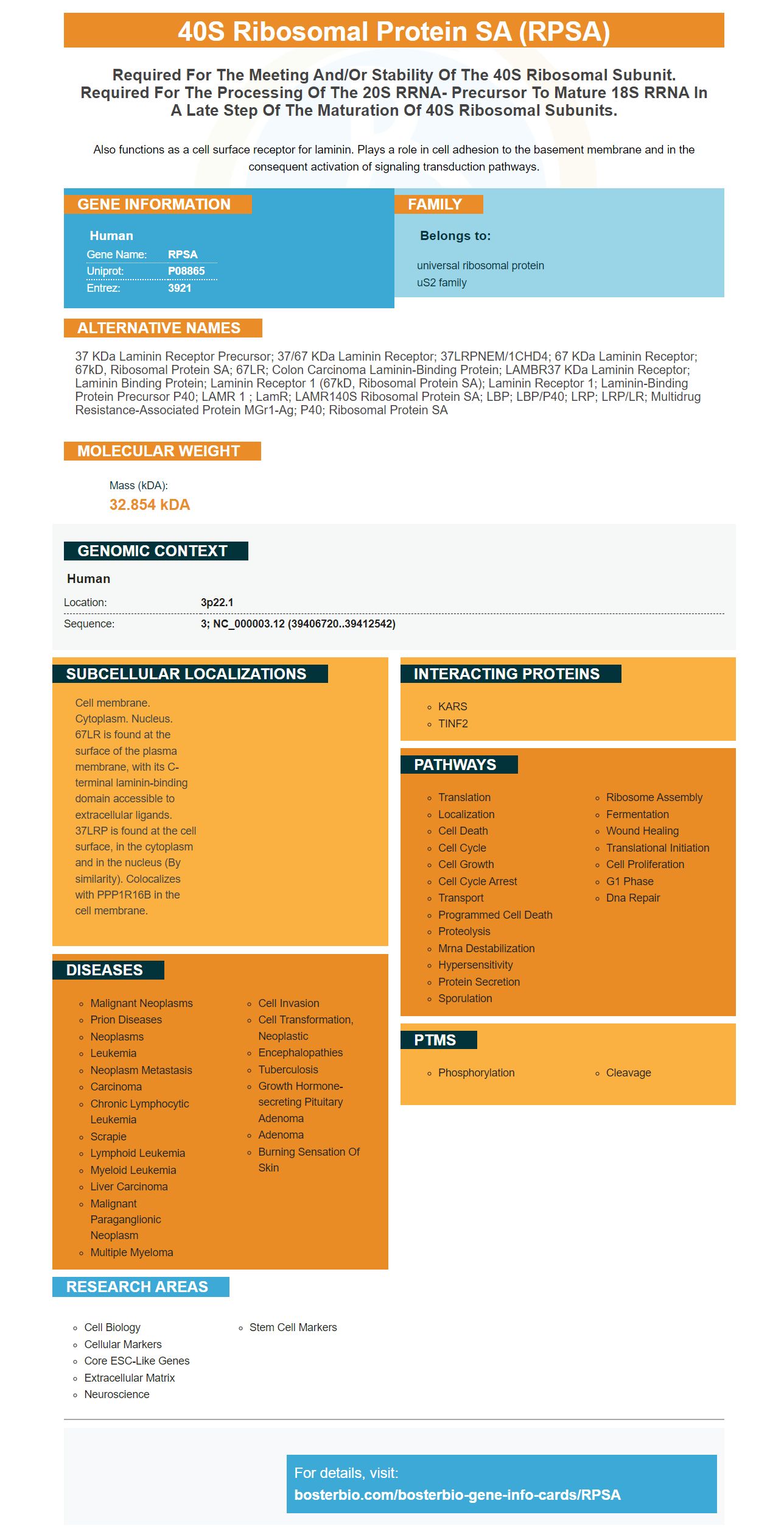
Facts about 40S ribosomal protein SA.
Also functions as a cell surface receptor for laminin. Plays a role in cell adhesion to the basement membrane and in the consequent activation of signaling transduction pathways.
Human | |
---|---|
Gene Name: | RPSA |
Uniprot: | P08865 |
Entrez: | 3921 |
Belongs to: |
---|
universal ribosomal protein uS2 family |
37 kDa laminin receptor precursor; 37/67 kDa laminin receptor; 37LRPNEM/1CHD4; 67 kDa laminin receptor; 67kD, ribosomal protein SA; 67LR; Colon carcinoma laminin-binding protein; LAMBR37 kDa laminin receptor; laminin binding protein; laminin receptor 1 (67kD, ribosomal protein SA); Laminin receptor 1; Laminin-binding protein precursor p40; LAMR 1 ; lamR; LAMR140S ribosomal protein SA; LBP; LBP/p40; LRP; LRP/LR; Multidrug resistance-associated protein MGr1-Ag; p40; ribosomal protein SA
Mass (kDA):
32.854 kDA
Human | |
---|---|
Location: | 3p22.1 |
Sequence: | 3; NC_000003.12 (39406720..39412542) |
Cell membrane. Cytoplasm. Nucleus. 67LR is found at the surface of the plasma membrane, with its C-terminal laminin-binding domain accessible to extracellular ligands. 37LRP is found at the cell surface, in the cytoplasm and in the nucleus (By similarity). Colocalizes with PPP1R16B in the cell membrane.
What are high-affinity primary antibodies (RPSA markers) and why is it important? Boster Bio: Let's take an overview of Steven Boster's background and RPSA marker. These antibodies have been around for more than 25 years. That means they're reliable, and they've been validated on Western Blotting, Immunohistochemistry, and ELISA.
The ribosomal protein SA is result of the RPSA gene. There are more than eighty different kinds of ribosomal protein in our cells. Each of them works together to implement the genetic instructions of cells to produce proteins. The ribosome's smallest subunit is home to the RPSA gene. The rpsA gene assists ribosomal proteins in processing the genetic instructions from cells to produce protein.
A complex mechanism of B-cell activation and T-cells are required to create high-affinity antibodies. The rPA marker was demonstrated to facilitate the selection of high-affinity cells by an affinity-driven process. Additionally, it provides an analysis of the gradual increase in the avidity of the antibodies stimulated by the GC response. It also helps to distinguish qualitatively between primary and secondary responses.
While there are many commercial sources of high-affinity primary antibodies however the quality of resulting antigen may differ. This can impact the outcome of your study. It is best to buy the Nb directly from the manufacturer to avoid these problems. This will ensure that the Nb source in the commercial market is managed. This method lets you obtain a high affinity primary antibody in only a few weeks.
RPSA-based antibodies can be used to identify a large number of microbes. They can also be used to identify biological substances and antigens that have high levels of cross-reactivity. High-affinity primary antibodies utilizing the RPSA marker can be utilized in clinical trials as a useful diagnostic tool. They are not made of light chains and are made of heavy chains. They are unique due to this.
The avidity of these antibodies was determined as the ratio between the binding rate and the dissociation rates. After the primary response, the avidity gradually increased. The peak of avidity remained stable during the first twenty days. After the boost, avidity increased fivefold. This was due to the increased dissociation rates and concentrations. These results indicate that RPSA-based methods can be extremely effective in discovering primary antibodies with high affinity.
Antibodies made using antigen-affinity purification are a great source of agents. These antibodies are highly specific and bind to target antigens efficiently and precisely. They can reduce background staining, which results in better reproducibility and higher productivity in research. These antibodies can also be used to identify antigens. Antigens can be identified in a myriad of applications by using the RPSA marker.
With the help of the rPA marker high-affinity primary antibodies can be created quickly. In animal models, it is recommended that you use SPR to evaluate the quality of rPA-specific antibody responses. This marker is a way to measure the quality of rPA specific antibodies found in polyclonal sera taken from mice that have been immunized experimentally. It is also helpful in the screening of antigen-specific antibodies in the human immune system.
These studies also demonstrated that the production of specific antibodies to rPA could occur earlier than monoclonal antibodies. This suggests that boosting with antigen can result in rapid increases in rPA specific antibodies levels and avidity, without the need for continuous GC development. Surface plasmon resonance-based plasma avidity measurements are another way to monitor humoral reactions. These results could aid in the development of immunotherapies and vaccines.
If you're looking for information on Steven Boster, you have come to the right place. Public records include Steve Boster's home address along with previous addresses and mobile phone numbers. Email addresses are also included. You can also determine if Steve Boster had any known relatives. You can narrow your search based on Steve's age or the location of his home. You can also get to know more about the background of Steve by reading his biography that written by him about himself.
PMID: 2970639 by Yow H., et al. Increased mRNA expression of a laminin-binding protein in human colon carcinoma: complete sequence of a full-length cDNA encoding the protein.
PMID: 2543954 by van den Ouweland A.M.W., et al. Characteristics of a multicopy gene family predominantly consisting of processed pseudogenes.
*More publications can be found for each product on its corresponding product page