This website uses cookies to ensure you get the best experience on our website.
- Table of Contents
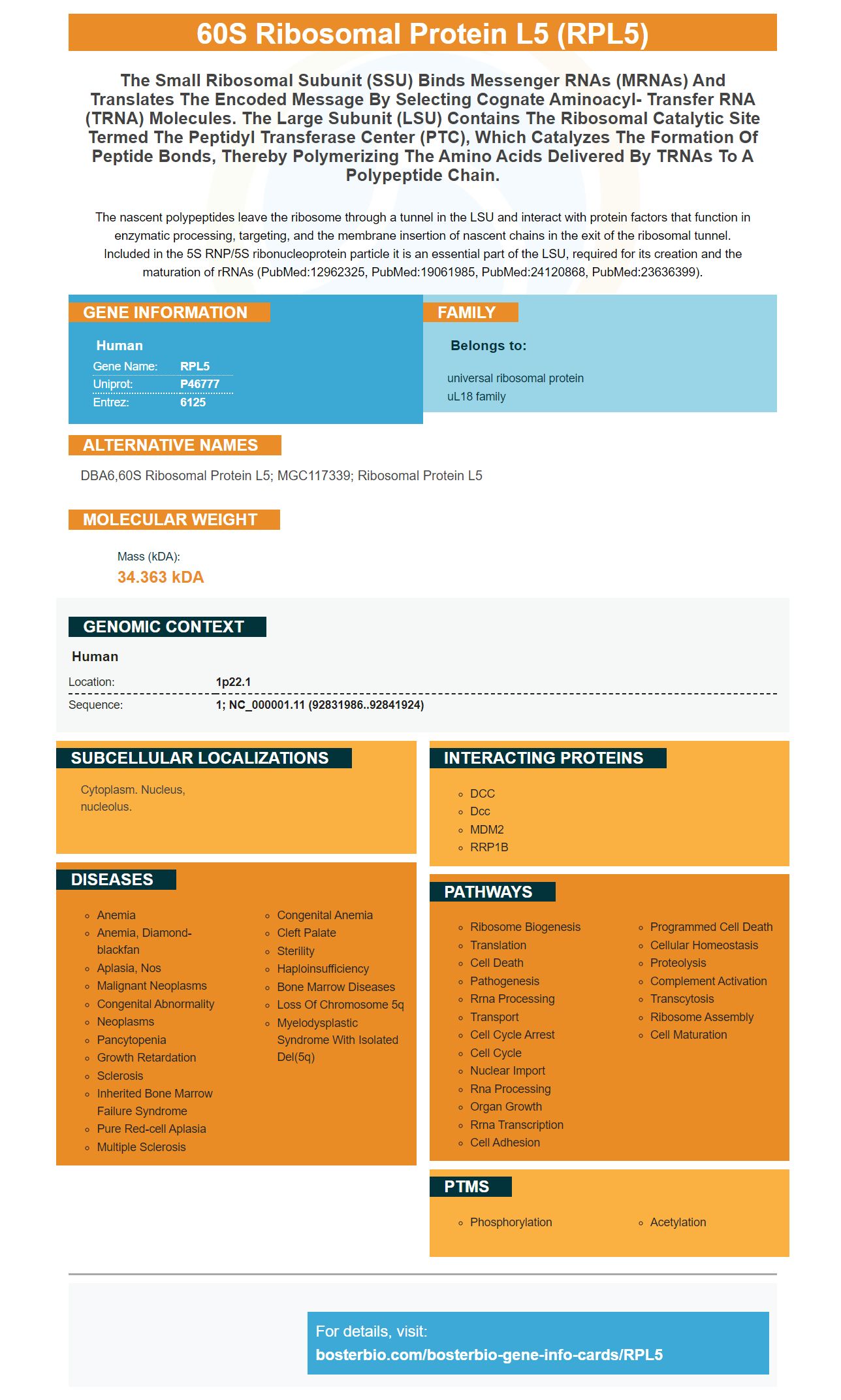
Facts about 60S ribosomal protein L5.
The nascent polypeptides leave the ribosome through a tunnel in the LSU and interact with protein factors that function in enzymatic processing, targeting, and the membrane insertion of nascent chains in the exit of the ribosomal tunnel. Included in the 5S RNP/5S ribonucleoprotein particle it is an essential part of the LSU, required for its creation and the maturation of rRNAs (PubMed:12962325, PubMed:19061985, PubMed:24120868, PubMed:23636399).
Human | |
---|---|
Gene Name: | RPL5 |
Uniprot: | P46777 |
Entrez: | 6125 |
Belongs to: |
---|
universal ribosomal protein uL18 family |
DBA6,60S ribosomal protein L5; MGC117339; ribosomal protein L5
Mass (kDA):
34.363 kDA
Human | |
---|---|
Location: | 1p22.1 |
Sequence: | 1; NC_000001.11 (92831986..92841924) |
Cytoplasm. Nucleus, nucleolus.
This article introduces Boster Bio, and Steven B. Boster (the company's president & chief scientific officer). It also provides information about RPL5/Ribosomal Protein L5 antibodies and the benefits of a high affinity primary antibody. For more information, please read the article titled "Antibody for Ribosomal Protein L5". It also explains why a high-affinity primary antibody is important for biological research.
The E. coli-expressed human RPL5 antibody has a His–Tag and a 1-297aa domain. The human RPL5 antibody is stable at room temperature and can be stored for one week at +2degC or 8degC. For longer storage, it should be stored at -20degC or -80degC. It should not be subject to repeated freeze-thaw cycles. Boster Bio is one of the leading suppliers of high-affinity, specific antibodies to RPL5.
Boster Bio has an RPL5 antiserum if you are looking for an antibody that is specific to Ribosomal Protein L5. Boster Bio's anti-RPL5 antibody (catalog no A02843-2), reacts with Human and has been successfully tested in both WB as ELISA. Because this antibody is used in multiple applications, dilution is an important consideration.
This antibody is designed for the detection of RPL5, also known to be MSTP030/PPP1R135/uL18. It has a mass of 34.4 kilograms and may have orthologs within yeast, plant and monkey. This description provides more information.
In a previous study, RPL5/Ribosomal Protein L5 antibody was used in order to determine the impact of a knockdown of c-Myc on metastasis. The antibody reduced the ability to human breast cancer cells invade and metastasize. The RPL5/Ribosomal Protein L5 monoclonal antiserum can be purchased.
This antibody recognizes NPM Protein, which is required to efficiently transport rpL5 through the nucleus and into the cell cytoplasm. It has been shown also to recognize NPM proteins, which are crucial for efficient transporting rpL5 out the nucleus. In addition to these activities, the Boster Bio Anti-RPL5/Ribosomal Protein L5 Antibody is also compatible with the NPM and g-tubulin proteins.
A high-affinity primary antibody has a unique epitope. It is here that the strongest binding occurs. This affinity determines the speed at which the antibody can bind antigens, the extent to the binding and the length of time it can bind. High affinity antibodies produce the best immune complexes. They are sensitive and quickly produce the most immune complexes.
If the antibody is highly immunogenic, it can bind both the RPL5 and its ligands. Antibodies with high affinity can cross-react with one another if they cross-react. If a single primary antibody is weakly binding but has high affinity, it will not detect the ligands. A high affinity primary antibody with high specificity for RPL5 is more effective at identifying antigens that have more than one antigen.
The DSaffinity fraction of mAbs was used in the identification and quantification of 107 proteins. Of these, 82 are located to the nucleus, while fifteen are associated with the mitotic cell cycle. These patterns could correspond to mitotic staining, as measured by the HEp-2 Test. The speckled patterns are likely to be caused by proteins associated with the cell cytoskeleton and vesicle.
A high-affinity primary antibody with multiple targets is better for mIHC, fluorescent, or mIHC detection systems. In contrast, a traditional chromogenic system relies on antibodies raised in different species or isotypes. This method is limited to three markers. Additionally, the primary antibody must be of different species and isotypes.
PMID: 7772601 by Frigerio J.-M., et al. Cloning, sequencing and expression of the L5, L21, L27a, L28, S5, S9, S10 and S29 human ribosomal protein mRNAs.
PMID: 12962325 by Odintsova T.I., et al. Characterization and analysis of posttranslational modifications of the human large cytoplasmic ribosomal subunit proteins by mass spectrometry and Edman sequencing.