This website uses cookies to ensure you get the best experience on our website.
- Table of Contents
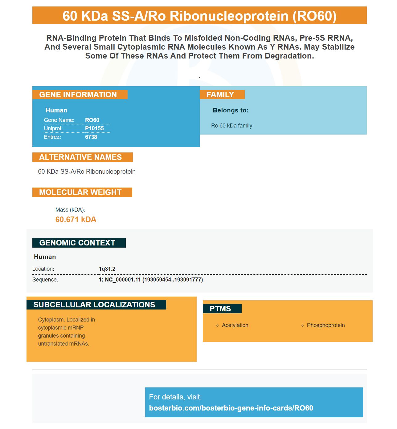


Facts about 60 kDa SS-A/Ro ribonucleoprotein.

.
| Human | |
|---|---|
| Gene Name: | RO60 |
| Uniprot: | P10155 |
| Entrez: | 6738 |

| Belongs to: |
|---|
| Ro 60 kDa family |

60 kDa SS-A/Ro ribonucleoprotein
Mass (kDA):
60.671 kDA

| Human | |
|---|---|
| Location: | 1q31.2 |
| Sequence: | 1; NC_000001.11 (193059454..193091777) |
Cytoplasm. Localized in cytoplasmic mRNP granules containing untranslated mRNAs.

When designing your next PCR experiment, it is important to understand how to use RO60 to stratify the antigen response. This intracellular antigen is 60 kDa in size and is used to stratify immune reactions to many molecules. RO60 is much more sensitive than other methods such as line immunoblots.
Ro60 is an intracellular, 60-kDa protein. It was initially described as a small complex of two proteins, Ro52 & Ro60, cytoplasmic nucleoproteins. Ro52 has been recognized as an individual antigen, due to new pathogenic mechanisms. Therefore, antibodies to Ro60 co-occur with those to Ro52.
There are several associations between Ro60 and SLE. SLE is strongly linked to anti-Ro60 and anti–Ro52/TRIM21. However, they are not associated with dsDNA, hypocomplementemia and Raynaud’s phenomenon. Both are important indicators of SLE. However, it is not clear if there are any associations between the antigens.
Recombinant SSA/Ro60 were attached to a sensor of a Biacore 3000 instrument. The concentration of the chip contained 1,000 molecules. The sensors recorded resonance in real time. This reflects the dynamic nature that the binding system is. The same preparation of antibody was used to measure both dissociation and association of the SSA/Ro60 combination.
Ro52 autoantibodies are specific to the p200 sequence of Ro52. Antibodies against the Ro52 protein correlate with AV delay in fetuses. The anti-Ro60 antiantigen correlates with heartblock in the same way. However, the mechanism for this association remains unclear. There is no consensus about the Ro60 autoantigen responsible for congenital heartblock. However, studies suggest that maternal antibodies directed towards a specific Ro52 sub-epitope, such as the p200, may be involved in the development of this condition.
Boster Bio, an American corporation based in Massachusetts developed the anti–Ro60 antibody. It was tested in mice cells in an animal model cutaneous leukemia. The anti-Ro60 antibodies stained human splenocytes when there was no Ro60 ligand. The anti–Ro60 antibody also reacted with the splenocytes in the wild-type C57BL/6 mouse.
The anti-SSA/Ro60 antibody showed mixed behaviours. Despite the lack of statistically significant differences between the two groups, anti-SSA/Ro60 antibody resulted significantly in lower haemoglobin in both positive and negative patients. The mean haemoglobin level in anti-SSA/Ro60-positive patients was within normal range. Platelet levels in positive patients were also significantly higher than in negative patients. This is surprising because anti-Ro60 antibody has been shown to decrease platelet production.
Anti-Ro60 is an intracellular antibody that forms a ribonucleoprotein compound with other noncoding RNA proteins, specifically the Y RNAs. The regulating of RNA and processing is crucially done by Y RNAs. Ro60 knockout mice show autoantibodies, glomerulonephritis, photosensitivity, and systemic lupus erythematosis-like syndrome. This illustrates the importance Ro60 & anti-Ro60 for the pathogenesis and progression of the disease.
Anti-Ro60, although a common serum antibody, is also useful for diagnostic purposes. However, most diagnostic laboratories report anti Ro60 results in qualitatively either positive or negative ways. This study was designed to determine the clinical significance of an antiRo60low subset identified in the past twelve months at a major international immunopathology diagnostic laboratory. Anti-Ro60low can be used as a way to categorize anti-Ro60low according to severity of disease.
In the study of RO60 versus anti-SSA/Ro52 antibodies, the RO60 marker exhibited a lower AUC at a cutoff of 14 U/ml than the other two methods. The RO52 marker had a significantly higher sensitivity at the same cutoff as the anti-SSA/Ro60. Although the markers have similar characteristics, the RO60 marker is more sensitive.
Sjogren's syndrome can be diagnosed with a variety of anti-Ro aabs. Ro60 is one example. It has been linked to anti-SSA and anti–TRIM21, which are proteins with similar molecular masses. Both are involved in protein ubiquitination as well as apoptosis. They are also part and parcel of a ribonucleoprotein protein complex that plays a key role in preventing the accumulation or misfolded of RNA. Anti-Ro60 is found in many autoimmune conditions and is associated several clinical manifestations.
The RO60 marker is more sensitive than other markers in detecting dsDNA. This marker was also sensitiver than anti-histone, anti-nucleosome. SLEDAI was also associated with anti-Ro52 antibodies and anti-nucleosome antibodies, but there were no significant correlations. It is important that the activity of SLE was associated with both RO60 and anti dsDNA antibodies, while the latter marker was not.
The IgM test is more sensitive in diagnosing neuroborreliosis than the IgG test. In an IgM test, the antibody response was 55% higher than the IgG in early stage disease. Similarly, Liu et al. found that the combined IgM and IgG response was 71% sensitive for early-stage samples of EM rash. Some people still question whether IgM and IgG tests are more sensitive than line immunoblots.
Regardless of the differences in the two methods, there are a few things that distinguish them. One is their simplicity and ease of use. The second is the possibility to perform automated interpretations. The line blot has a similar sensitivity to the ELISA. However, it is less sensitive. It is faster to perform and takes much less time to process. It is, however, more sensitive than line immunoblots.
Specificity and sensitivity are often not easily comparable. Some studies defined a positive result as either IgM or IgG, and report the sensitivity based on one test while others use both. However, in some studies, both IgM and IgG negatives showed higher sensitivity. A few studies used a definition for a positive result that was based on the presence or absence of certain antibodies. This is especially problematic for clinical specimens, which could contain weak antibodies and samples from patients with weakened immune system.
Specificity and sensitivity are closely related. The results of a test determine how sensitive it is. The higher the test's sensitivity, the greater the chance of false-positive results. Specificity and sensitivity are affected by the incubation time for ELISA and Western blot testing. The test's sensitivities can vary depending on how an antibody was isolated. If you are concerned about the test's sensitivity, consider a different test.
PMID: 3200833 by Deutscher S.L., et al. Molecular analysis of the 60-kDa human Ro ribonucleoprotein.
PMID: 2649513 by Ben-Chetrit E., et al. Isolation and characterization of a cDNA clone encoding the 60-kD component of the human SS-A/Ro ribonucleoprotein autoantigen.


