This website uses cookies to ensure you get the best experience on our website.
- Table of Contents
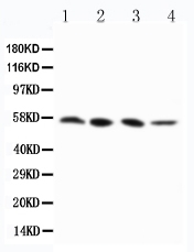
Facts about Receptor-interacting serine/threonine-protein kinase 3.
RIPK3 binds to and enhances the action of three metabolic enzymes: GLUL, GLUD1, and PYGL. These metabolic enzymes can eventually stimulate the tricarboxylic acid cycle and oxidative phosphorylation, which could result in enhanced ROS production.
Human | |
---|---|
Gene Name: | RIPK3 |
Uniprot: | Q9Y572 |
Entrez: | 11035 |
Belongs to: |
---|
protein kinase superfamily |
EC 2.7.11.1; Receptor-interacting protein 3; receptor-interacting serine/threonine-protein kinase 3; receptor-interacting serine-threonine kinase 3; RIP3; RIP-3; RIP3receptor interacting protein 3; RIPK3; RIP-like protein kinase 3
Mass (kDA):
56.887 kDA
Human | |
---|---|
Location: | 14q12 |
Sequence: | 14; NC_000014.9 (24336025..24339991, complement) |
Highly expressed in the pancreas. Detected at lower levels in heart, placenta, lung and kidney. Isoform 3 is significantly increased in colon and lung cancers.
Cytoplasm, cytosol. Cell membrane. Mitochondrion.
In this article, we'll discuss the BosterBioIL6 marker as well as its uses. This marker reduces the growth and tumorigenicity in prostate cancer cells. It also enhances three metabolic enzymes. It is degraded by CHIP. This marker is useful in monitoring the effectiveness antimicrobials. But what are its best uses, you ask? Continue reading for more information.
It is well-known that RIPK3 plays a crucial role in the regulation of the activation of RIPK3 pathways. Its activities include modulating necroptosis and inflammasome activation. It also controls mitochondrial function, cellular metabolic activities, and other functions. Moreover, RIPK3-associated injuries do not necessarily cause high levels of cell deaths. Genetic deletion studies have shown that RIPK3 may play a non-cell-death role in this process.
Although RHIM is a heterodimer, it is not enough to trigger necroptosis. RIPK3 homo-oligomerization is required for activation, phosphorylation, and recruitment of MLKL. RIPK1 deletion also triggers RIPK3–MLKL-mediated necropsis. It is not known if RIPK1 deletion actually enhances RIPK3 oligomerization.
Caspaxe-8 is mediated by RIPK1 (RIPK3). Both RIPK1-RIPK3 are key mediators in necroptosis. The inhibition of caspase8 enhances necroptosis. RIPK3 promotes necroptosis when it is overexpressed. It also initiates apoptosis.
HFD was not increased in mice with RIPK3 knockouts. The mice showed basic glucose intolerance. However, HFD exacerbated inflammation, steatosis, and apoptosis, which are diametrically opposed to alcohol. And RIPK3 knockout mice developed more inflammatory-dependent hepatitis than those that had normal livers.
Recent genetic experiments show that RIPK3 plays a critical function in regulating inflammatory responses in the TNF-receptor pathway. RIPK3 is also important in the regulation of epithelial integrity. In a mouse model of Gaucher’s disease, RIPK3 expression has been shown to be associated with better survival. Further, RIPK3 deficiency also increases motor coordination and reduces cerebral injury.
RIPK3 activates caspase-8dependent apoptosis. RIPK3 activates NLRP3-dependent inflammasome. This is independent of RIPK1. These results support the idea that RIPK3 could be a valuable drug target. RIPKL insufficient mice can lead to necroptosis. This is a condition that causes embryonic lethality and liver inflammation.
Recent research shows that MMAC/PTEN suppresses cells' proliferative capacity. This inhibition was independent of p53 mutation. MMAC/PTEN also decreased the size of prostate cancer cells and prolonged their survival, suggesting that MMAC/PTEN might be a therapeutic target. However, more studies are needed to fully understand the mechanisms underlying MMAC/PTEN-mediated tumorigenesis.
In mice, ex-vivo treatment of PC3 cells with MMAC/PTEN decreased their tumorigenicity and cellular proliferation. These cells had a reduced tumorigenicity, and were less likely than others to invade lymph nodes. This inhibition is likely due the MMAC/PTEN-mediated control of various signaling pathways, factors, and factors released by prostate carcinoma cells. Continue reading to learn more about MMAC/PTEN.
DT-13 has antitumor activity. DT-13 also suppresses PI3K/Akt signalsing and inhibits PDK1 Akt. It also prevents the growth of prostate cancer cells by inhibiting their metastasis. It also inhibits the growth PC3 and DU145 cell. However, it blocks the p70S6K/mTOR-axis.
Genetically engineered mice suggest that MMAC/PTEN may play some role in the development and early stages of prostate tumors. Embryonic lethality can be caused by the loss of either MMAC/PTEN genes. MMAC/PTEN+/ mice can also develop various types of tumors including lymphoid hyperplasia and autoimmune syndrome. These findings suggest that this phenotype might be due to the loss of one or more of these alleles.
In a study of DT-13 in vitro, this compound was found to inhibit the proliferation of PC3 and DU145 cells. Cell colonies were treated at 0, 2.5, 10 mM and 48 h respectively. The size of the cells was determined by a flow cytometer. These data represent three independent experiments. It inhibits the growth of prostate cancer cells through inducing death in these cells.
Chinese academy sciences provided the human prostate cancer cells. They were cultured using RPMI 1640 medium containing 10% fetal bifid serum from Biological Industries. The tumors also were fixed in formalin, embedded in paraffin and then removed. Histological examinations revealed whether the cells were metastatic. There were no interactions between Ad-GFP or adenosine.
Researchers examined whether RIPK3 increases the activity three metabolic enzymes involved with cell death in a UUO mouse model for kidney fibrosis. The results showed that RIPK3/ mice had fewer TUNEL-positive kidney cells at day 7 than controls. However, the UUO-treated mice had a lower number of TUNEL positive cells, while RIPK3-/ mice had fewer TUNEL+ cells than WT.
RIPK3 has two important functions. It can induce apoptosis, but not necroptosis, in certain situations. It is known to activate and recruit caspases by phosphorylating its evolutionary conserved threonine residues. However, the exact mechanism behind RIPK3 activated caspases varies depending on the cell type.
In aging female mice, the RIPK3 marker phosphorylation occurs at S164/T165. This mutation is not critical for female reproductive function as corpus luteum development occurs during the ovulation cycle. Mutant mice with S165a/T166A mutations display hyper-ovulation. These mice were not sexually mature so RIPK3 phosphorylation shouldn't affect their reproductive function.
RIPK3 also acts as a transcriptional regulator for cFLIP. This gene regulates basal self-phagy and instigates increased autophagosome production. Mutations at the serine 164 site can cause this activation. However, knocking out cFLIP does not block RIPK3-induced autophagosome formation. However, it enhances its activity for basal autophagy.
The RIPK3 molecule has pleiotropic ER stress effects. It regulates transcriptional response to metabolic stresses by dampening proinflammatory and anti-oxidant genes. RIPK3 inhibition prevents b-cell loss. These findings require further research to confirm. It is important not to forget that the ER can be a key upstream component of metabolic stress.
The RIPK3 protein is involved in innate immunity. RIPK3 activates apoptosis related proteins such as TRAIL, TNF receptors, and death receptors. In addition, RIPK3 promotes the activity of three metabolic enzymes, which results in increased ROS production. RIPK3 can be considered a marker for innate immunity.
RIPK3 plays a critical role in necroptosis regulation and is negatively regulated through CHIP, an E3 ligase. However, RIPK3 was expressed more in cells lacking CHIP and were more sensitive for TNF-induced nephrotoxicosis. These results suggest RIPK3 may be a useful target to treat cancer.
CHIP is also implicated as an OGD protective factor. It plays multiple functions in protein quality control, including cochaperone function as well as ubiquitin-ligase activity. The discordance in RIPK3 mRNA levels and protein levels suggests that post-transcriptional mechanisms might be involved in protein degradation. To confirm this, the mutant CHIP gene was transfected into N2a cells.
Recent research suggests that anisomycin may inhibit eIF2a phosphorylation. This could help to protect against ischemic stroke. In an endoplasmic reticulum stress situation, CHIP was also overexpressed and abrogated eIF2-a phosphorylation. Further research on this protein could lead to the development of drugs that treat necroptosis. Understanding the mechanisms by which RIPK3 is controlled and controlled by CHIP may help to develop better treatments.
CHIP binds the Hsp70 proteins, which ubiquitinates RIPK3 at K55 and K89 in human cells. Inhibiting CHIP can lead to the induction and maintenance of poly-Ub links at these three sites. This mechanism is involved with necroptosis. CHIP is also implicated with human diseases, such as Alzheimer's.
CHIP is a negative regulator of RIPK1 and RIPK3 necrosome formation. It acts as a desensitizing factor to TNF-mediated necroptosis. The CHIP protein is a crucial regulator of RIPK3 function. It promotes mTOR expression. CHIP may regulate RIPK3 expression in the intestinal tract.
In the mouse model, knockout mice lacking RIPK3 had more advanced colon cancer than wild-type (WT) mice. Tsc1 IEC deficiency led to a significant increase colon cancer development, in WT mice. However the effect was 1.7 times less in Ripk3+/ animals. Furthermore, tumors in Tsc1IEC-KO/RIPK3-KO mice had lower necroptosis and were associated with a greater number of large tumors than WT mice.
PMID: 10339433 by Yu P.W., et al. Identification of RIP3, a RIP-like kinase that activates apoptosis and NFkappaB.
PMID: 10358032 by Sun X., et al. RIP3, a novel apoptosis-inducing kinase.
*More publications can be found for each product on its corresponding product page