This website uses cookies to ensure you get the best experience on our website.
- Table of Contents
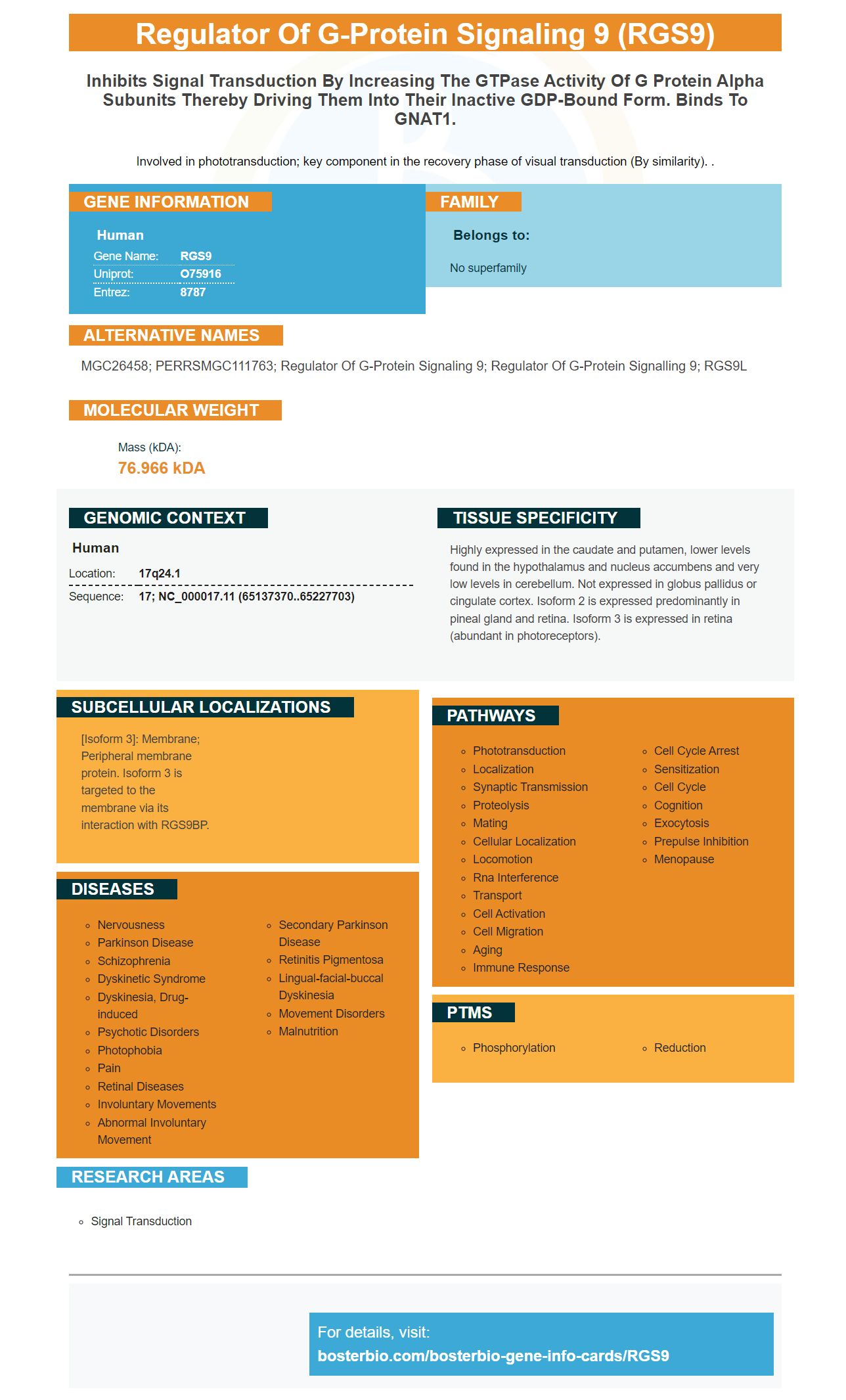
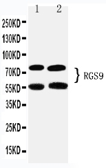
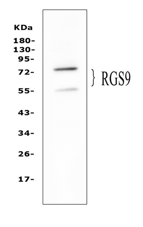
Facts about Regulator of G-protein signaling 9.
Involved in phototransduction; key component in the recovery phase of visual transduction (By similarity). .
Human | |
---|---|
Gene Name: | RGS9 |
Uniprot: | O75916 |
Entrez: | 8787 |
Belongs to: |
---|
No superfamily |
MGC26458; PERRSMGC111763; regulator of G-protein signaling 9; regulator of G-protein signalling 9; RGS9L
Mass (kDA):
76.966 kDA
Human | |
---|---|
Location: | 17q24.1 |
Sequence: | 17; NC_000017.11 (65137370..65227703) |
Highly expressed in the caudate and putamen, lower levels found in the hypothalamus and nucleus accumbens and very low levels in cerebellum. Not expressed in globus pallidus or cingulate cortex. Isoform 2 is expressed predominantly in pineal gland and retina. Isoform 3 is expressed in retina (abundant in photoreceptors).
[Isoform 3]: Membrane; Peripheral membrane protein. Isoform 3 is targeted to the membrane via its interaction with RGS9BP.
The RGS9 indicator is a commonly used biomarker in many biological assays. The antibody, either monoclonal or polyclonal, reacts with RGS9 in a variety of animal samples. Boster Bio has used rabbit and mouse as models for the development of antibodies to detect RGS9. This biomarker plays an important role in phototransduction, particularly during the recovery phase of visual transduction.
The RGS9-2 subcellular protein has a distinct distribution pattern. It is detected by Western blot after sequential Triton X-100 elutions. An RGS9-2 primary antibody will yield high-affinity antibodies that recognize RGS9 marks. This study used an RGS9-2 monoclonal immunoglobulin to detect RGS9-2 within brain sections from wildtype mice.
The immunoreactivity of RGS9-2 is seen in puncta along the dendritic shafts of 14 DIV striatal-cortical co-cultures. Further, we observed that RGS9-2 and PSD-95 staining overlapped. This revealed corticostriatal spines and synapses. As a result, RGS9-2 immunoreactivity in cultured rat brains was extremely efficient.
Recombinant Fab can be generated in parallel and is highly automated, allowing for multiple projects to be completed at once. An affinity tag attached to a high-affinity antibody can capture beads, and allow you to complete many projects simultaneously. It should be noted that not all peptides yield high-affinity recombinant antibodies; further studies are needed to determine the optimal target properties.
Quantitative immunoblotting of isolated nuclei, detergent soluble homogenates, or unfractionated start material has been performed. The samples were transferred onto supported nitrocellulose and immunoreactivity was measured using HRP, enhanced chemiluminescence, and LI-COR IR secondary antibodies. The data presented are mean values of three measurements. The results of these experiments show the sensitivity of this method but are not conclusive.
The RGS9-2 Protein has a unique 25kD C-terminus. This makes it difficult in vivo for the nuclear locization machinery to access it. It is possible that RGS9-2 can interact with other signaling proteins, such as the Drd2 or M1 muscarinic Acetylcholine receptors. These studies are ongoing.
The KD distribution for RabMAb was compared to that for mouse MAb. The affinity of the rabbit antibodies was higher than that of monoclonal mouse antibodies. The OIRD measurements were taken at UC Davis and compared with a statistical model. The results showed that the two methods were strongly correlated. KD values indicate the affinity and sensitivity for the antibody.
Molecular biology studies have revealed that the RGS9 gene is largely expressed outside of the retina. It appears to have a specific effect on neuronal pathways in brain. Moreover, the RGS9 protein is expressed in neurons in various regions of the body, including the striatum, the nucleus accumbens, and the neostriatum. RGS9's mRNA is also found in brain's nucleus and olfactory tubes.
One study found that mice with an RGS9-knockout were weighed before they were given each drug. San Diego Instruments supplied a photobeam cage and rack activity system. The animals were allowed 30 minutes to become used the light beam before being given drugs. Baseline activity measured the concentrations of the drugs in animals before administration. After the animals had been conditioned the drugs were administered intraperitoneally to them.
The effects of acute morphine and chronic morphine on RGS9 levels have been studied in the spinal column and thestriatum. This latter region is crucial for opiate analgesia and reward. Morphine-resistant RGS9 mutant mice have delayed tolerance to the drug. However, chronic morphine administration did not reverse the effects of the drugs on the spinal cord. The RGS9 Gene Marker is a promising tool that can be used to identify the genetic basis of opioid sensitivity.
In this study, the motor behavior was not affected by 6-hydroxydopamine lesions. This suggests that multiple feed-back mechanisms are part of the basal glandular circuitry. For example, amphetamine caused a 1000 fold increase of dopamine in the synapses when administered. By identifying the role of RGS9 in this circuitry, the researchers hope to develop drug compounds that target this receptor.
RGS9 interacts in mouse models with dopamine D2 receptors. To study this interaction, researchers created mice bearing a mutation in the RGS9 gene. RGS9-cre knockout is a mouse method that allows the generation of mice with the RGS9 mutation. These mice can be fertile and live. The RGS9 gene-knockout mice are created when the striatum is deleted from the loxP-flanked DNA sequence.
RGS9 expression levels in adult brain tissue are very low. Previous studies have identified RGS11, RGS9 proteins in embryonic mouse brains and retinas. These markers aren’t specific for NPCs. It is therefore impossible to determine if RGS9 gene expression is specific or not for neural stem cells. However, it might help distinguish NPCs apart by detecting their DNA. To understand the role RGS9 plays in differentiating NPCs we need to first determine its source.
GGL-containing RGS Proteins always exist in complexes, in vivo. GPB-2 functions are unlikely to be affected by RGS9 unless the cell has been infected. In addition, GPB-2 is involved in regulating the level of phosphorylation in the brain. The RGS9 marker is still being discovered. These are key points that help to define the function and purpose of this marker.
The gene is expressed in deep areas of the brain, including the striatum. It appears to be involved signaling pathways involving the chemical messenger dopamine. Dopamine is responsible for planning, coordinating and coordinating movement. It may also play a part in the brain's response to opioid drugs. It is also found in the cerebellum which controls the endocannabinoid systems. RGS9 is a key player in drug addiction.
The RGS9 gene encodes two proteins, RGS9-1 and RGS9-2. The RGS domain includes flanking sequences, and is found throughout the entire cell's genome. RGS9 functions through its flanking sequences. Although the functions of these proteins are different, they have similar functions. RGS9 is expressed within the brain in adult animals. This information will assist researchers in determining which RGS9 variations are present within brain tissue.
PMID: 9765512 by Granneman J.G., et al. Molecular characterization of human and rat RGS9L, a novel splice variant enriched in dopamine target regions, and chromosomal localization of the RGS9 gene.
PMID: 10564809 by Zhang K., et al. Structure, alternative splicing, and expression of the human RGS9 gene.