This website uses cookies to ensure you get the best experience on our website.
- Table of Contents
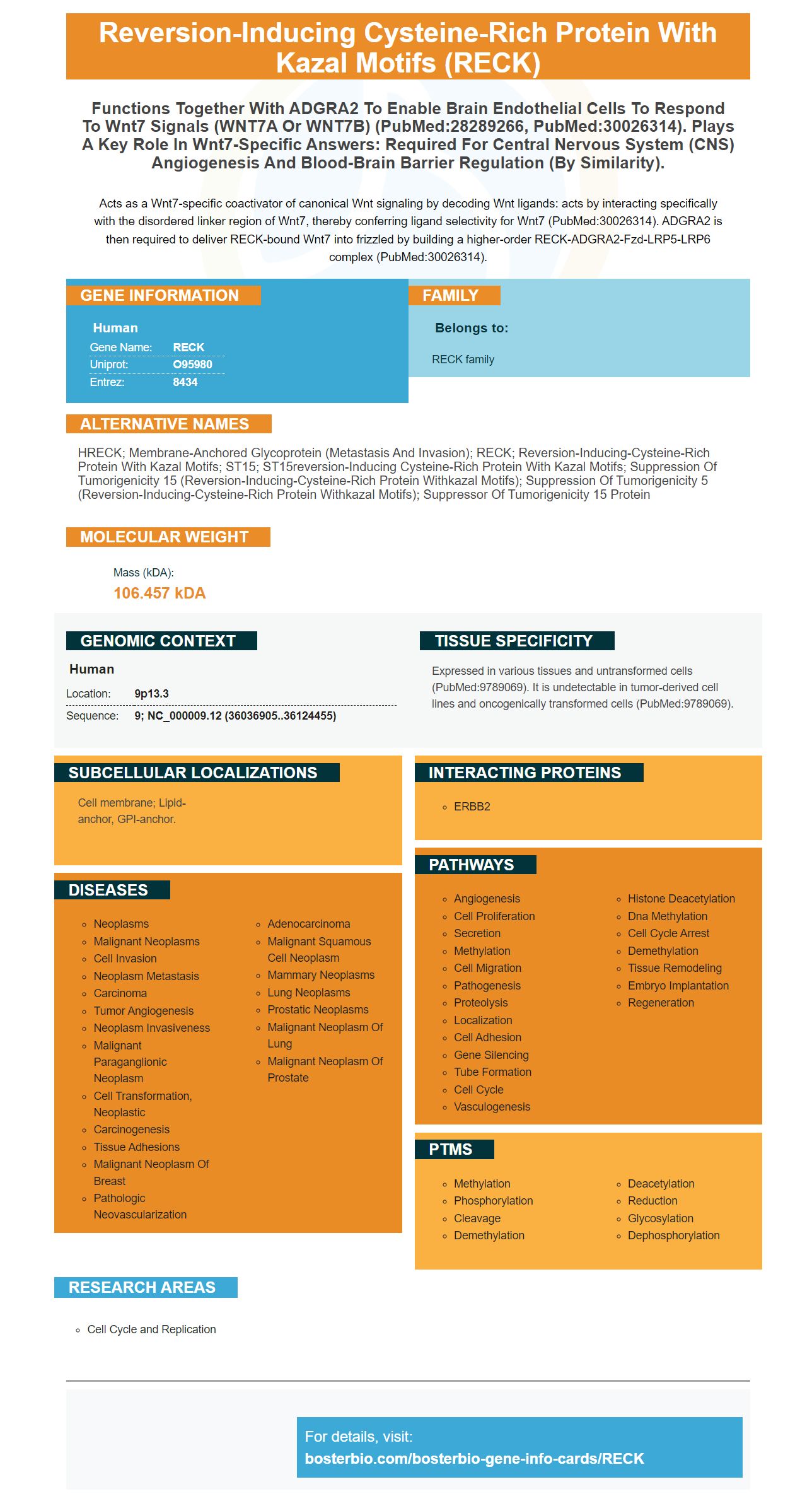
Facts about Reversion-inducing cysteine-rich protein with Kazal motifs.
Acts as a Wnt7-specific coactivator of canonical Wnt signaling by decoding Wnt ligands: acts by interacting specifically with the disordered linker region of Wnt7, thereby conferring ligand selectivity for Wnt7 (PubMed:30026314). ADGRA2 is then required to deliver RECK-bound Wnt7 into frizzled by building a higher-order RECK-ADGRA2-Fzd-LRP5-LRP6 complex (PubMed:30026314).
Human | |
---|---|
Gene Name: | RECK |
Uniprot: | O95980 |
Entrez: | 8434 |
Belongs to: |
---|
RECK family |
hRECK; membrane-anchored glycoprotein (metastasis and invasion); RECK; reversion-inducing-cysteine-rich protein with kazal motifs; ST15; ST15reversion-inducing cysteine-rich protein with Kazal motifs; suppression of tumorigenicity 15 (reversion-inducing-cysteine-rich protein withkazal motifs); suppression of tumorigenicity 5 (reversion-inducing-cysteine-rich protein withkazal motifs); Suppressor of tumorigenicity 15 protein
Mass (kDA):
106.457 kDA
Human | |
---|---|
Location: | 9p13.3 |
Sequence: | 9; NC_000009.12 (36036905..36124455) |
Expressed in various tissues and untransformed cells (PubMed:9789069). It is undetectable in tumor-derived cell lines and oncogenically transformed cells (PubMed:9789069).
Cell membrane; Lipid-anchor, GPI-anchor.
The RECK marker was developed in 1993 by Steven Boster, who gave his company the nickname, "He who converts science in the lavatory." He has been involved in the development of hundreds of antibodies, including several for use in immunohistochemistry (IHC). By the late 90s, he had become the largest catalog antibody company in China. PicoKine is Boster’s proprietary ELISA platform which delivers high-sensitivity ELISA kits for researchers.
The RECK Marker (which stands for Reactive Element Capture) is used to detect proteins during Western blot experiments. The RECK marker can be purchased in a variety color-coding schemes including red, green and blue, white, yellow, and white. This versatile marker can be used for immunohistochemistry, western blotting, and ELISA. Boster Bio: Best Uses of The RECK marker
Different antibody detection methods can be used for the detection of antibodies in sample gels. For example, the colorimetric technique uses a colored precipitate as a way to detect a signal. A chemluminescence-based method uses a light-emitting compound to detect an antibody. A fluorescence method uses an antibody that has been fluorescently marked with a fluorophore.
ECL and chromogenic detection are the two main methods for western-blot antibody detection. Both of these methods depend upon the substances bound the secondary antibodies. Enzymatic labels require additional steps but are extremely sensitive. Commonly, alkalinephosphatase as well as horseradish peoxidase can be used. Although alkaline phosphatase offers many advantages over other enzymes it can also have a slower reaction time and a higher background signal which can lead to a low signal–to-noise ratio.
Western blotting (or protein immunoblotting) is a widely used analytical technique for the detection and quantification of target proteins within samples. This method involves the separation of protein samples by polypeptide length using SDS-polyacrylamide gel electrophoresis. The proteins are then transferred onto a membrane. After staining, the proteins are visualized using antibodies specific for the target protein antigens. The intensity of a specific reaction is used for identification.
Many of the Western blot antibodies from Boster Bio are specific for the ubiquitinated CHAF1A. The anti-HA antibody detects CHAF1A's ubiquitinated version. The results of Western blot analysis can be interpreted and used to help with research in various areas. It is important to make sure that the Western blot method of detecting antibodies works correctly.
Protein transfer efficiency is determined by the staining of membranes with Ponceau S (0.02% w/v) or 5% glacial acetic acid. After incubation, the membrane is washed three times in TBS Wash Buffer to remove unbound antibodies. A second stain, which can be used to detect a primary antibody is applied to your membrane and incubated overnight.
An error in detection can be caused by the use of the wrong antibodies or a poor sample. Sometimes, the blot might show bands that are too faint and not enough signal. You can solve some problems by using another sample, or changing the running buffer. Alternativly, you might need to heat the sample to break down the quaternary structure. In this case, a high voltage level and air bubbles may lead to a band with uneven width.
A chemiluminescent system uses light to detect proteins in membranes. This two-component method is composed of a stable peroxide and an enhanced substrate solution. It is typically a luminol based HRP. Both components are mixed together in equal amounts. The result is light with a wavelength 425 nm. This light can be detected by X-ray films. CCD camera imagers cooled by cooling provide faster data capture and greater sensitivity.
Amersham ECL Prime is a highly sensitive reagent, allowing for multiple exposures in the same run. Because it has a high-intensity signal, it is compatible with multiple and different blots. Hybond ECL membranes consist of reinforced nitrogencellulose and provide excellent sensitivity. The ECL Plex Western Blotting System can detect all sizes of proteins.
The optimal conditions for light generation in western blotting can last from six to 24 hours. The substrate used and the enzyme ratio are key factors in the duration of light production. The peak light emission occurs during the first five- to thirty minutes of incubation. Researchers can optimize the parameters of their experiments using the signal emission curve of the chemiluminescent detection device to achieve reproducible results.
The ECL chemiluminescent detector kit includes a luminol based chemiluminescent sub-substrate that detects horseradish oxidase. It is compatible with several blocking buffers. It can also use nitrocellulose membranes. Because it has a low background, it is compatible with various WesternBright systems and substrates. The ECL chemiluminescent detection device can visualize a blot in low background. It can be viewed on CCD imaging systems and x-ray film.
ECL chemiluminescent detection systems are popular for protein detection in western blots. A HRP-labeled second antibody is combined with a horseradish peoxidase enzyme. This reaction creates light at a wavelength (428 nm), which is detectable using an Xray film or digital imager. If light is unavailable, a photochemical examination can be done.
DAB chromogenic detector system is an Intensification tool that significantly increases chromogenic signals from immunohistochemistry. It uses horseradish (HRP) as the catalyst to deposit two separate Linkers. These Linkers are then detected using streptavidin-peroxidase conjugate and diaminobenzidine (DAB).
PMID: 9789069 by Takahashi C., et al. Regulation of matrix metalloproteinase-9 and inhibition of tumor invasion by the membrane-anchored glycoprotein RECK.
PMID: 18194466 by Chang C.K., et al. The Kazal motifs of RECK protein inhibit MMP-9 secretion and activity and reduce metastasis of lung cancer cells in vitro and in vivo.
*More publications can be found for each product on its corresponding product page