This website uses cookies to ensure you get the best experience on our website.
- Table of Contents
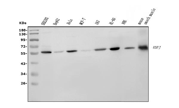
Facts about Recombining binding protein suppressor of hairless.
When associated with some NICD product of Notch proteins (Notch intracellular domain), it functions as a transcriptional activator that activates transcription of Notch target genes. Likely represses or activates transcription via the recruitment of chromatin remodeling complexes containing histone deacetylase or histone acetylase proteins, respectively.
Human | |
---|---|
Gene Name: | RBPJ |
Uniprot: | Q06330 |
Entrez: | 3516 |
Belongs to: |
---|
Su(H) family |
CBF1; CBF-1; CSL; H-2K binding factor-2; IGKJRB1; IGKJRB1RBPSUHCBF1; IGKJRBRBP-J kappa; immunoglobulin kappa J region recombination signal binding protein 1; J kappa-recombination signal-binding protein; KBF2; RBPJ; RBP-JK; RBPJKMGC61669; RBP-Jrecombining binding protein suppressor of hairless (Drosophila); RBPSUH; recombination signal binding protein for immunoglobulin kappa J region; recombining binding protein suppressor of hairless; Renal carcinoma antigen NY-REN-30; SUH; suppressor of hairless homolog
Mass (kDA):
55.637 kDA
Human | |
---|---|
Location: | 4p15.2 |
Sequence: | 4; NC_000004.12 (26163458..26435131) |
Nucleus. Cytoplasm. Mainly nuclear, upon interaction with RITA/C12orf52, translocates to the cytoplasm, down-regulating the Notch signaling pathway.
Using the RBPJ marker in your research will increase your knowledge about how to test the effects of chemicals on your cells. Luckily, the RBPJ marker is a highly versatile tool. Several different methods are available, such as gel electrophoresis, western blot, and other techniques. This article will highlight the benefits of RBPJ marker and its uses.
Boster Bio offers antibodies that react to the RBPJ marker in Mouse, Rat and Human tissues. The company uses monoclonal antibodies with high affinity and is cited extensively in the scientific literature. The antibodies are validated on ELISA, Immunohistochemistry, and Western Blotting. The Boster Bio RBPJ markers are available for purchase online. If you are an educator and want to use the Boster Bio RBPJ markers, you may use these products to teach your students about this protein and its function in human cells.
To use the RBPJ marker for immunofluorescence staining, you must first incubate cells in test tubes or microtiter plates with fluorescently-conjugated antibodies. Then, you will use a flow cytometer machine to analyze the staining results. The RBPJ antibody is the ideal choice for detecting these proteins in a variety of studies.
The RBPJ gene is a crucial component of the T reg cell that regulates immune function. The deletion of this gene reduces the differentiation potential of T reg cells, causing these cells to overexpress Gata-3 and other T H-2 associated proteins. Therefore, the antibody that targets this gene is essential to distinguish sick from healthy T cells. This research also provides new insights into the role of Rbpj in the immune system and the mechanisms that regulate its function.
RBPJ binds most strongly at peaks near the borders of nucleosomes. Binding is preferentially centered on the peaks of both the same and opposite strands. The RBPJ binding peak scores and nucleosome coverage are plotted as a fraction of the overall distribution. The highest nucleosome coverage and scores were observed near the peak summits of RBPJ and at positions close to the 5' end of bound nucleosomes.
Besides the SRF binding motif, RBPJ also binds to methylated Alu elements. The RBPJ binding peak summits were found to be co-localized with positive regulatory marks, including Alus and SRF. Moreover, 59% of the non-Alu subset showed high H3K9 acetylation and RNA Pol II binding. This indicates that RBPJ and SRF factors are intimately linked in regulating gene expression.
The Rabbit Anti-RBPJ antibody recognizes human, bovine and canine RBPJ. RBPJ is a transcription factor that targets notch signaling. It is also implicated in suppression of differentiation in myogenic precursor cells. High-affinity Rabbit Anti-RBPJ antibody targets the RBPJ marker. This antibody is a unique monoclonal antigen, specifically designed to recognize RBPJ.
These gene infographics based on the RBAJ marker show the distribution of the gene trkA, trkB, and trkC in the DRG of Rbpj-deficient mice. Each arrow points to the corresponding DRG, and each bar represents 100 m. Gene infographics based on the RBPJ marker further describe gene expression.
The role of RBPJ in the DRG is not yet clear. It has been suggested that Rbpj inhibits neuronal differentiation of DRG NCCs by repressing NeuroD1 expression. NeuroD1 has a role in both promoting neurogenesis and suppressing gliogenesis, although the exact mechanisms involved in defective gliogenesis in CKO mice remain unclear.
The RBPJ Marker is a key component of the Notch pathway and regulates the cellular response to membrane-bound ligands. Notch2 regulates endothelial cell proliferation, migration, and angiogenesis and acts as a receptor for membrane-bound ligands. Boster Bio's RBPJ markers are available in various lengths to fit your needs.
This Mouse RBP4/Retinol Binding Protein ELISA kit contains 96 wells and removable strips for easy and accurate quantitation of Mouse Rbp4 in various samples. It is compatible with a variety of sample types including serum, urine, EDTA, and cell culture supernatants. Its sensitivity is 10 pg/ml and there is no detectable cross-reactivity between the Mouse Rbp4 protein and other samples.
Several studies have shown that blocking the Notch signaling pathway improved myocardial injury in mice. The Notch pathway also promotes M2 polarization in cardiac macrophages and reduces fibrosis. Using the Boster Bio RBPJ ELISA kit, researchers were able to identify various macrophage types in the myocardial infarct area of mice. They were also able to measure HYP content and COL1 expression.
PMID: 8406481 by Amakawa R., et al. Human Jk recombination signal binding protein gene (IGKJRB): comparison with its mouse homologue.
PMID: 8016657 by Henkel T., et al. Mediation of Epstein-Barr virus EBNA2 transactivation by recombination signal-binding protein J kappa.