This website uses cookies to ensure you get the best experience on our website.
- Table of Contents
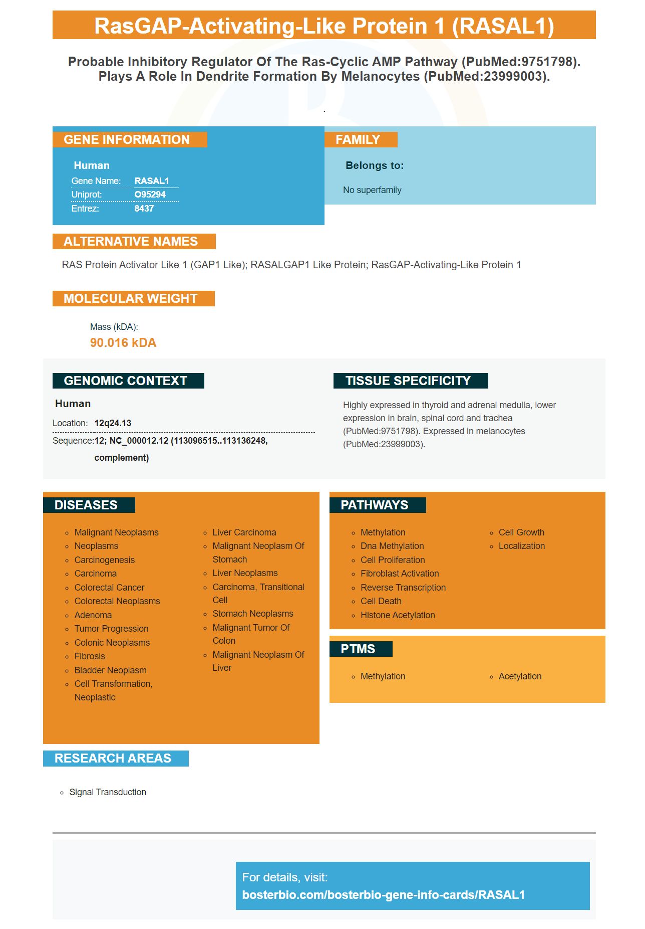
Facts about RasGAP-activating-like protein 1.
.
Human | |
---|---|
Gene Name: | RASAL1 |
Uniprot: | O95294 |
Entrez: | 8437 |
Belongs to: |
---|
No superfamily |
RAS protein activator like 1 (GAP1 like); RASALGAP1 like protein; rasGAP-activating-like protein 1
Mass (kDA):
90.016 kDA
Human | |
---|---|
Location: | 12q24.13 |
Sequence: | 12; NC_000012.12 (113096515..113136248, complement) |
Highly expressed in thyroid and adrenal medulla, lower expression in brain, spinal cord and trachea (PubMed:9751798). Expressed in melanocytes (PubMed:23999003).
This article will focus on the use of Boster Bio’s Anti-RASAL1 marker in Immunofluorescence and Transfection. Both are necessary for high-quality antibodies. Read on to learn about these applications and how to get started. We will also discuss the differences between commercial and Boster Bio anti–RASAL1 markers.
The PicobandTM catalog includes the Boster Bio Anti-RASAL1/Rasal1Marker. This antibody reacts with Human and Mouse proteins. It is also available in liquid form in 50% Glycerol. It has been tested in Western Blotting and Immunohistochemistry. For more information, please visit the manufacturer’s website.
Communication and epithelial cell adhesion are crucial for cancer progression. Epithelial cells undergoing EMT typically express mesenchymal cells and increase RASAL2 expression. In addition, RASAL2 has been implicated in cancer metastasis and EMT. Boster Bio Anti-RASAL1/RASAL1 Marker is therefore available from many manufacturers and has a wide range.
RASAL2 regulates the epithelial-mesenchymal transition. It regulates ERK, p-Raf-1 and other proto-oncogenes. It promotes EMT in CRC cells. RASAL2 knockdown caused an increase in expression of pRaf-1 as well as ERK. Moreover, RASAL2 inhibits the activity of the epithelial-mesenchymal transition in human CRC cells.
Boster Bio Anti-RASAL1/RasAL1 markers can detect RASAL2. It was tested in CRC cell lines and a wild type colon line (NCM460). The results showed that RASAL2 expression was significantly lower in CRC tissues. Its decreased level was also associated to a decrease in protein levels. RASAL2 is therefore downregulated in CRC tissues.
The RASAL1 marker first became known in the late 1990s. It is expressed in cancer cells. It is crucial for the development of the disease. It is expressed in various cell types, including breast cancer and hepatocellular carcinoma. Although it isn't clear why this marker exists, it is believed to play a role at EMT, which is an important step in cancer progression.
Steven Boster originally developed this antibody in 1993. This product has been a commercial success across many areas. It is currently one among the most well-known antibodies. It has also been used for cell invasion, migration and adhesion experiments. This antibody is highly sensitive and is available in a variety of formats, including ELISA, western blotting, immunohistochemistry, and more.
Recent research showed that RASAL2 gene expression was significantly lower in CRC cell lines than in untransfected cells. This could indicate that RASAL1 may be downregulated within CRC tissues. Further studies are required to determine if RASAL1 -shRNA can be used to treat colorectal tumors. What are the best ways to use the RASAL1 genetic marker?
In addition to its role in epithelial-mesenchymal transition, RASAL2 regulates p-Raf-1 expression, ERK, and N-cadherin. Western blotting allowed us to determine the protein levels. This marker can help to determine if the protein's presence is necessary for cancer development. If it is, it is important that you understand how RASAL1 affects transition from epithelial into mesenchymal.
Boster Bio offers antibodies to the RASAL1 Marker. These antibodies are produced on boster bio's proprietary Picoband(tm) platform and have been extensively validated in Western Blotting, Immunohistochemistry, and ELISA. The Boster Antibody binds to RASAL1 and can be used in a wide range of biological samples. Boster Bio Antibodies are designed for use in a variety of experiments and are highly cited in the research community.
Besides its role in the epithelial-mesenchymal transition, RASAL1 also regulates the expression of Raf-1, a proto-oncogene. The protein p-Raf-1 is phosphorylated and ERK, E-cadherin, and vimentin was assessed with reverse transcription-quantitative polymerase chain reaction.
RASAL2 is essential for EMT during tumor progression. Epithelial cells expressing RASAL2 are able to migrate and divide into mesenchymal cells. ERK-activated RASAL2 facilitates tumor progression via EMT and metastasis. However, little research has been done on this marker in CRC. Its expression could be related to the development of the cancer.
It is also used in colorectal cancer. A loss-of function RASAL2 cell model was established. Additional studies are currently underway to determine its role. Furthermore, RASAL2 protein expression is compared to levels in wild-type colorectal cell lines. These preliminary results show that RASAL2 could be a therapeutic target for CRC.
The RASAL1 marker may play an important role in liver cancer development. This marker was identified for the first time by genome-wide analyses of methylation. Experiments with RASAL1-knockout mice and in vitro studies showed that hypermethylation of RASAL1's promoter suppressed its expression. Hyperactivated Ras was associated to increased type I collagen, intrinsic proliferative activity, and a-SMA. The methyl-CpG bindin protein was also found to regulate RASAL1 in rat liverfibrosis.
As a result of the activation of the Ras-MAPK pathway, the RASAL1 marker suppresses the activity of HSCs. As expected, RASAL1 suppresses HSC activity. It is not known how RASAL1 suppresses HSC activation. This protein plays multiple roles in the liver. Its expression is highly controlled by SMA.
Although the RASAL1 gene marker has many applications for cancer diagnosis, it remains controversial. We examined the ability of the RASAL1 test to identify patients with advanced liver carcinoma. RASAL1 is downregulated in liver cancer tissues and may be a reliable predictor of survival. This marker inhibits HIF-2a via the ERK1/2 pathway, which may affect the proliferation of tumor cells. This study offers a new direction in clinical use.
It has been demonstrated that RASAL1 plays a significant role in HIF-1 and HIF-2a regulation. RASAL1 inhibits HIF-2a and restores gluconeogenic gene transcription in Vhlh mutant mice. It also prevents hypoglycemia. HIF-2a also plays a critical role in liver cancer metabolism. In the meantime, research is underway to explore how the RASAL1/HIF-2a axis could become a therapeutic target for liver cancer.
The RASAL1 tumor suppressor has been linked to colorectal and bladder carcinomas. It is not clear what molecular mechanisms are behind RASAL1 tumor suppression. These tumors show that RASAL1 might play a role for the development of fibrisis. It could also play a role in the development atypical colorectal carcinomas.
PMID: 9751798 by Allen M., et al. Restricted tissue expression pattern of a novel human rasGAP-related gene and its murine ortholog.
PMID: 23999003 by Yoo J.C., et al. SYT14L, especially its C2 domain, is involved in regulating melanocyte differentiation.