This website uses cookies to ensure you get the best experience on our website.
- Table of Contents
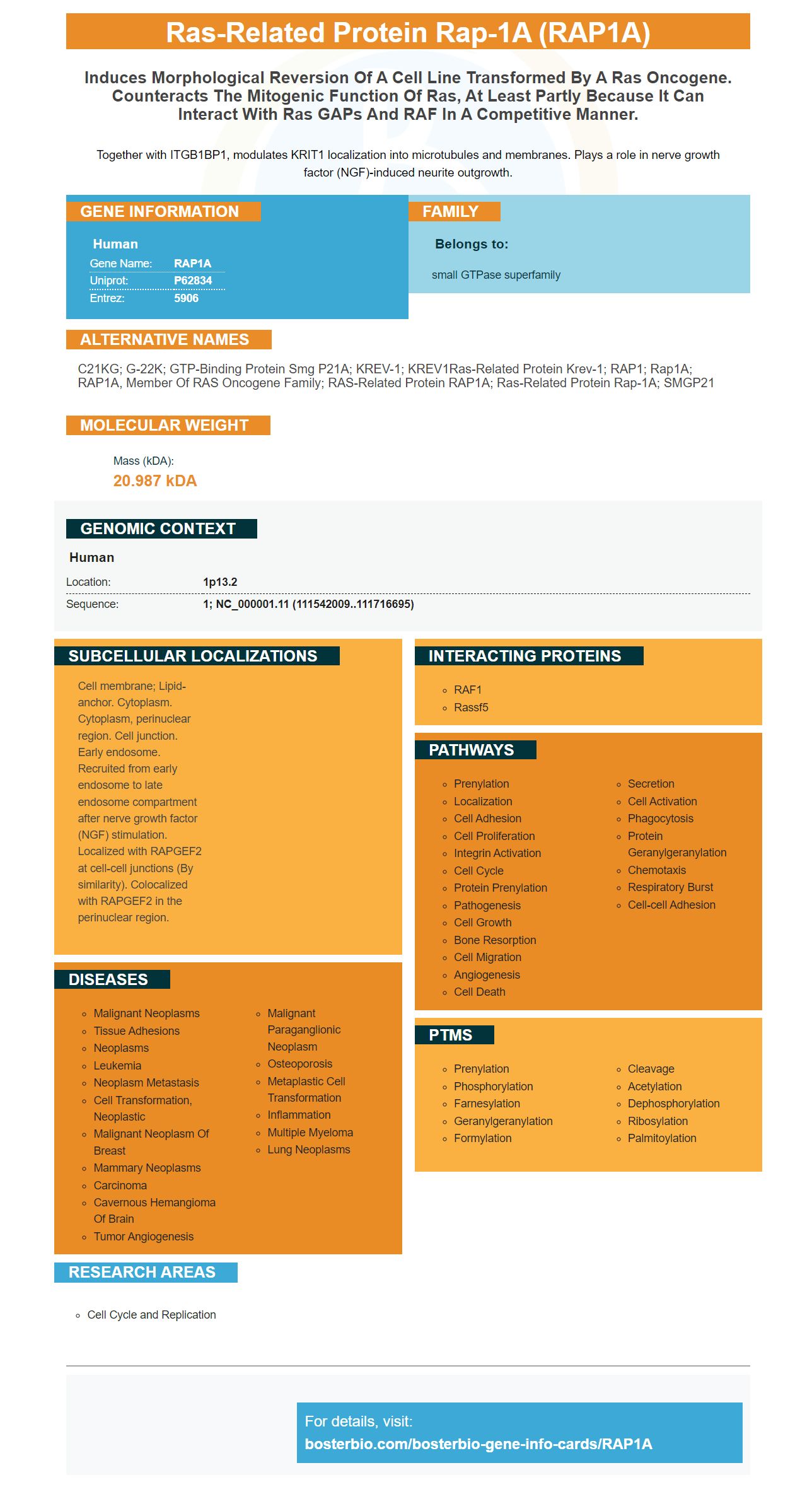
Facts about Ras-related protein Rap-1A.
Together with ITGB1BP1, modulates KRIT1 localization into microtubules and membranes. Plays a role in nerve growth factor (NGF)-induced neurite outgrowth.
Human | |
---|---|
Gene Name: | RAP1A |
Uniprot: | P62834 |
Entrez: | 5906 |
Belongs to: |
---|
small GTPase superfamily |
C21KG; G-22K; GTP-binding protein smg p21A; KREV-1; KREV1Ras-related protein Krev-1; RAP1; Rap1A; RAP1A, member of RAS oncogene family; RAS-related protein RAP1A; ras-related protein Rap-1A; SMGP21
Mass (kDA):
20.987 kDA
Human | |
---|---|
Location: | 1p13.2 |
Sequence: | 1; NC_000001.11 (111542009..111716695) |
Cell membrane; Lipid-anchor. Cytoplasm. Cytoplasm, perinuclear region. Cell junction. Early endosome. Recruited from early endosome to late endosome compartment after nerve growth factor (NGF) stimulation. Localized with RAPGEF2 at cell-cell junctions (By similarity). Colocalized with RAPGEF2 in the perinuclear region.
This article will show you how MAPKRAP1A signaling in HCC can be linked to a better prognosis. This article also discusses the LASSO method, which is used in developing a prognostic sign. It will also talk about its clinical applications. We'll also discuss the best uses for this marker. This information can be used by all scientists around world.
Metastasis from primary cancers to the peritoneum can be controlled by the CX3CL1/ICAM-1 feedback loop between VBMECs, NSCLC cell and VBMEC cells. NSCLC tumors are prevented from spreading to spinal metastases by inhibiting this signaling network. It prolongs the life expectancy of mice with NSCLC.
MAPK-RAP1A signaling plays a key role in cancer progression, as well as being a tumor's master regulator. The risk factor for developing hepatocellular carcinoma is known to be the upregulation of this signaling pathway. MAPK/RAP1A signaling is also associated with immune invasion and tumor size.
This study also investigated the function of KSR2 within HCC cells. KSR2-knockdown cell lines grew smaller tumors when compared to KSR2-overexpression cells. Furthermore, the tumors with higher KSR2 levels were associated with a poor prognosis and a shorter OS.
KSR2 is an important scaffold within the MAPK-RAP1A pathway. KSR2 is an important scaffold in the MAPK-RAP1A pathway. High KSR2 levels decrease HCC cells' sensitivity for sorafenib. Furthermore, it is possible to inhibit KSR2 through targeted therapies such as the kinase inhibitor sorafenib.
CX3CR1 inhibition can have a beneficial impact on osteolytic bones lesions. CX3CR1 knockdown significantly reduced bone lesions. CX3CR1 KD treatment resulted in significant reductions in MMP-2 levels. The inhibition of CX3CR1 has been shown to inhibit tumor cell growth.
This study shows that mice with CX3CR1-knockout mutations live longer than those with LFA-1 mutations. Therefore, the findings of this study suggest that the therapy may improve the treatment of HCC. Two phase-III studies support the study. The authors identified an inhibitor for CX3CR1 as well as another one that targets NF-kB.
Researchers discovered that RPS8 can be used as a biomarker for alcohol related HCC. The Cancer Genome Atlas Database (TCGA), contained gene data profiles. The researchers then selected the best genes and performed gene coexpression network analysis. They identified three hub genes within the protein-protein interactions network that were significantly related to child-Pugh score (T-stage), and bodyweight.
The LASSO algorithm was used to create a gene signature. It was then tested for prognostic validity. The resulting gene Signature had a statistical significance score of 0.897 and a positive predictive power score of 1.088. The TCGA/BRCA cohort database was used to assess the gene signature. To determine the risk score of BC-patients, we ran a Cox regression with a least absolute selection and operator (LASSO) penalized algorithm. Our gene signature was also compared to an independent validation set, GSE7390.
The 17-gene signature helped us classify patients as high- or low-risk. Then, we tested the prognostic ability of the new protein signature by comparing actual survival with predicted survival. The results of this study are summarized in Table 1.
The LASSO algorithm was used to develop a multigene risk signature using the TCGA-BRCA cohort. The genetic expression level of the genes was determined through GO pathway, KEGG pathway, and DAVID (6.8) analyses. Next, we applied the LASSO penalized Cox regression algorithm to extract genes that were prognostic related. We then performed multifactorial Cox regression analysis for the genes identified by the LASSO algorithm and used the coefficients of the genes in each gene to construct the risk score equation.
The LASSO algorithm is a unique genetic risk signature that has been validated to be used for EC by the miR-93-2p inhibitor. It has a strong prognostic ability and can predict response to immunotherapy and chemotherapy. Despite these strengths the signature is still a highly promising prognostic risk indicator for EC. The validation cohort validated its prognostic significance.
The study also evaluated the expression of nine protein levels related to MSI status and tumor grade. The protein signature had a positive effect on prognosis in EC patients, according to the researchers. This signature was found to accurately predict survival rates in this group of patients. In addition to improving prognostic scores the results revealed that the 11 Gene Model may be an effective biomarker for predicting risk in EC patients.
The LASSO algorithm is an integral component of the cyclic coordinate descent algorithms. It has several advantages over the other algorithms. The entire sequence used in creating the model is the default value. The lasso algorithm is nonlinear and the objective function replaces the observed response values with their weighted or prior values. A shrinkage penalty can also be applied to variables.
The LASSO is a regression method that uses regularization and variable choice. These techniques improve predictability and interpretability of statistical models. Robert Tibshirani invented the term "lasso". This method of regression analysis is very versatile. Multicollinearity is solved by the R language. It can be used in order to find the minimum value for a variable.
The LASSO algorithm often is used in predicting model parameters. This method can be used in both simple and complex models. The data values of the model are reduced to a central point. This method performs L1 regularization by adding a penalty equal the absolute coefficients. It can also automate variable selection or parameter elimination. The LASSO algorithm is an ideal choice for multicollinear models because it can reduce parameter and variable selection.
The LASSO method is a statistical technique which forces the coefficients into zero. It works in a similar way to the ridge regression but with fewer coefficients. Its inputs are b 0 and b1, b2,..., bp. The LASSO algorithm uses c (combined) function of R language. You can modify the parameters and the degree to which regularization are used to customize the algorithm.
When you perform a LASSO algorithm in R language, it's important to note that two covariates are identical for each observation. The LASSO objective function is the sum of two covariates whose coefficients are the same. The objective of a laso algorithm is the first thing that you need to understand. The most common method for data analysis is the LASSO algorithm. This algorithm works well on datasets containing large amounts of data and a variety of features.
The RAP1A protein is one the most abundant in the human genome. But what does it do? It is involved in DNA repair, and the regulation of gene expression. The inability to repair double-strand breaks via NHEJ is reduced by the loss of RAP1. RAP1A levels were also found to predict the efficacy for chemotherapy in breast and colon cancers.
Rap1 may also play an important role in stem cells of other types, including hematopoietic. Increased levels Rap1 have been linked to increased vascularpermeability. Recognizing patients with this gene is critical for early detection of CVD. However, more research is needed in order to determine if Rap1 plays an important part in vascular permeability. The clinical applications for the RAP1A marker are discussed below.
Rap1A is a multifunctional protein that may be related to context-dependent expression. This protein can be found in the hypothalamus. It is a key location in the CNS responsible for energy and glucose metabolism. Rap1 is a key player in metabolic processes. This has been confirmed by genetics, pharmacology and glucose clamp studies. It is believed RAP1A may be involved in insulin signaling, and glucose metabolism.
RAP1A has a role in the repair/replacement of damaged chromatin. It is also associated to the NHEJ complex. RAP1A, when activated by the cytokines is responsible for the formation this complex. When activated, RAP1 also binds DNA, thereby activating the NFkB signaling pathway. RAP1A plays an important role in NHEJ's DNA repair.
In vivo, Rap1 regulates NHEJ. Rap1KO mice had a greater tumor size and an acceleration in tumorigenesis. Rap1KO mice also had increased B-cell populations. The B cells were increased by g-H2AX staining and immunohistochemistry analysis revealed increased cell proliferation. Rap1KO mice also had higher g–H2AX stains in their spleens.
Rap1 is involved in many other functions in mammals, including tumor growth. Rap1A acts as a marker for tumor-related activities. The gene can be used for many clinical purposes, including aging, gastrointestinal disease and metabolic disorders. It can also be used to identify a patient's age, race, gender, and overall health. It is important to note that RAP1A has multiple roles in mammals, including growth, reproduction, and immune response.
PMID: 3045729 by Pizon V., et al. Human cDNAs rap1 and rap2 homologous to the Drosophila gene Dras3 encode proteins closely related to ras in the 'effector' region.
PMID: 2507536 by Nagata K., et al. Purification, identification, and characterization of two GTP-binding proteins with molecular weights of 25,000 and 21,000 in human platelet cytosol. One is the rap1/smg21/Krev-1 protein and the other is a novel GTP-binding protein.