This website uses cookies to ensure you get the best experience on our website.
- Table of Contents
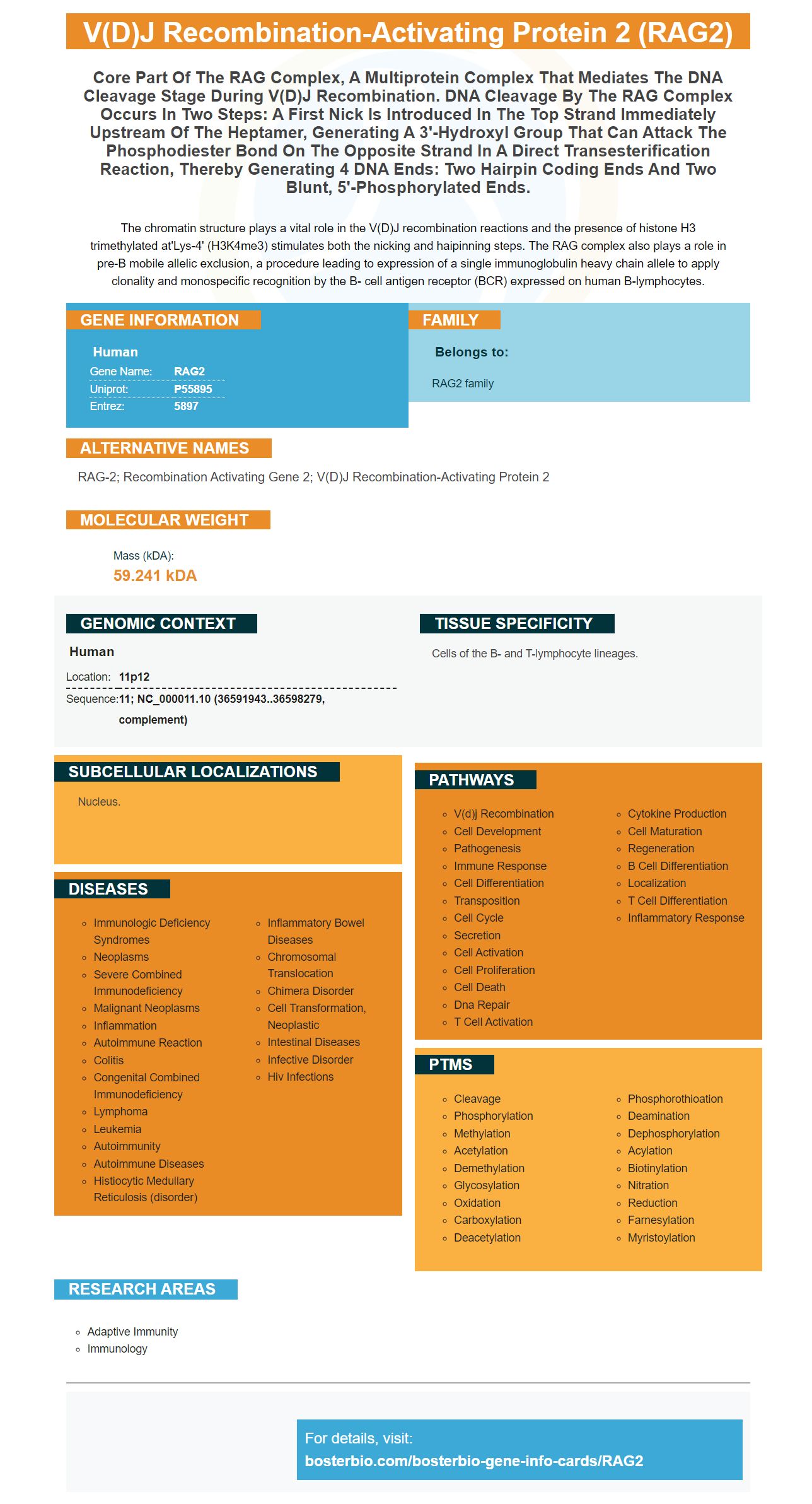
Facts about V(D)J recombination-activating protein 2.
The chromatin structure plays a vital role in the V(D)J recombination reactions and the presence of histone H3 trimethylated at'Lys-4' (H3K4me3) stimulates both the nicking and haipinning steps. The RAG complex also plays a role in pre-B mobile allelic exclusion, a procedure leading to expression of a single immunoglobulin heavy chain allele to apply clonality and monospecific recognition by the B- cell antigen receptor (BCR) expressed on human B-lymphocytes.
Human | |
---|---|
Gene Name: | RAG2 |
Uniprot: | P55895 |
Entrez: | 5897 |
Belongs to: |
---|
RAG2 family |
RAG-2; recombination activating gene 2; V(D)J recombination-activating protein 2
Mass (kDA):
59.241 kDA
Human | |
---|---|
Location: | 11p12 |
Sequence: | 11; NC_000011.10 (36591943..36598279, complement) |
Cells of the B- and T-lymphocyte lineages.
Nucleus.
This article will cover how RAG1 bonds to RSS and how RAG2 recruits RAG into H3K4me3. It will also discuss how this binding leads to DNA distortion. Continue reading to learn more about how to use the RAG2 marker for boster bio. We hope that this article was helpful. Good luck! We hope that this article was as enjoyable as it was for you!
The RAG Complex catalyzes nicking (strand formation) and cleavage (strand cleavage). It binds both to 12 and 23-RSS substrates and introduces an nick at a junction of a coding segment. The RAG Complex also recruits proteins from classical nonhomologous-end joining (NHEJ). These proteins join and process coding segments. High-mobility group proteins act as cofactors of RAG complexes, and their recruitment promotes RAG activity and RSS bending.
RAG1 binds to RSS only if RAG1 binds to RAG2. These proteins amplify the signals produced by DNA cleavage and are bound to single-stranded molecules of DNA. RAG1 can bind multiple DNA molecules but it is limited in its ability to distinguish between specific targets and non-specific ones. It may not be relevant whether RAG1 binds specifically to DNA molecules or RSS in cells.
RAG1 binding to RSS can cause conformational changes for both RAG1 monomers. RAG1 binding might allow for the formation of a pair with the complementary 12-RSS/23RSS. RAG1 can be bound to RSS to induce catalytically efficient conformations in RAG1, like the E984 of a23. Further research is needed to determine the exact mechanism of RSS binding in human cell cells.
In vitro, cells can co-exist with RAG1 or RAG2 In vitro studies of RAG2 in cells have demonstrated that the two proteins induce circle formation, but the resulting circles are less than expected. Similar research on RAG1 ligation revealed that RAG1-D708A/RAG2-DNA compounds increased circle formation by two. This suggests that higher RAG1 amounts promote HMG2 sequestration. This could be a factor in preventing circle formation.
The structures of RAG1/DNA complexes offer a glimpse of RSS recognition within the heptamer. The SEC and PC structure of the RAG1/RAG2 complex exhibit almost identical RSS interactions. These structures were determined with a symmetrized SEC. The bound RSS from both proteins can be clearly seen at the first fifteen positions in the heptamer. These positions form a region directly contacting RAG.
A RAG2 complex, which binds to the same substrates as RAG1 and RAG2, was also discovered. RAG1 alone and RAG1 plus RAG2 both bind to the same signal with the same efficiency. These complexes then underwent a series if binding reactions. Later, it was discovered that these RAG proteins are sequence specific when they bind the same substrate.
In the RAG1-DNA binding assay, 25fmol of labeled sub-DNA was mixed with 0.25pmol of unlabeled competitive DNA. The RAG1-DNA complex remained stable for more than an hour after addition of competitor DNA. The RAG1–RAG2-DNA complex was highly stable. Prior to gel electrophoresis, the RAG1-1-DNA complex was reduced half-way by competitor DNA. The RAG1-1-RAG2-DNA compound remained stable for at the most for about an hour.
The RAG complex plays an important role in the recombination process of V(D),J loci in the context of immunodeficiency. These loci contain hypermethylated histones H3K4me3, which is a key feature during the recombination process. Its interactions with H3K4me3 is essential for the development and function lymphocytes. The RAG complex plays an important role in the regulation V(D)J Recombination.
Two mechanisms are involved when the RAG complex is recruited to the site where the DSB is located. The other involves the recruitment of DNA damage response factor ATM. It acts transiently upon the second allele and repositions them to pericentromeric hemochromatin (PCH). During repair the RAG binding to the uncleaved alele is depleted by its transient relocation towards the PCH.
The interaction of RAG2 with PHD was determined by using peptides containing H3K4me3 motifs. The peptides had monomethylated, asymetrically methylated, and symmetrically-methylated R2. These peptides contain protein-peptide complexes. The mutant protein binds R2-methylated H3 peptides with decreased affinity. This could be due to its W453-destabilized structure.
RAG is believed to be responsible for nucleosome remodelling, including the H3K4me3 area. RAG proteins may play a role in DNA recombination. RAG2 is also located in the nucleus' euchromatic regions. Both RAG proteins play a critical role in gene expression. For gene expression regulation to take place, it is crucial that there is a balance between transcription factors as well as remodeling factors.
The RAG2 Protein recognizes methylated Histone H3 at arginine one and lysine patru. This recognition triggers translocation, activation of V(D),J RAG complex. Recent studies have also indicated a role for the regulation of gene transcription by methylating arginine. If this role is emphasized, it may lead to the discovery of a new gene regulatory pathway.
RAG plays a key role in V(D]J recombination. This is due to its association with highly conserved RSS at V(D]J loci. These loci have thousands of RSS like sequences and are likely mistargeted. T-ALL demonstrates all the hallmark features of VDJ-recombination. It also shows the presence of translocation between cryptic RSS sites.
In addition to recruiting RAG to H3K4me3 regions, RAG2 can also initiate V(D)J recombination in lymphocytes. This can lead to the creation of diverse antigen receptors. This recombination happens in a specific lineage. In addition, chromatin structure is critical for V(D)J loci. Hypermethylation at lysine4 of histone H3 is a prerequisite for RAG to trigger V(D)J recombination. This is when the RAG1/RAG2 complex recruits RAG H3K4me3 to initiate double-strand breaks at RSS websites. To initiate V(D),J Recombination, the RSS website must be in an Open Chromin Context
The Thr490 methylation residues in the noncore region can also impact RAG2's localization and dynamics within the pre-Bcell nucleus. These differences may have implications on activation of IgK-recombination in this particular cancer type. This research shows that Rag2 recruits RAG2 H3K4me3 for tumor suppression in patients with Rag2 mutations.
RAG1-RAG2 binding results in significant DNA distortion. To be biologically significant, the target site must have sufficient affinity and avidity to distinguish it from non-specific DNA. RAG1 fails this test due to its lack of sufficient affinity. Similar observations were made regarding RAG2 binding. Here are two examples of how this alteration occurs. In both cases, the distortions are significant.
RAG1 & RAG2 cleave DNA in a similar manner to the cleavage process, but the proteins interact with the VDJ recombination signal. Complex formation requires a divalent ion. In addition, RAG1 and RAG2 bind DNA with greater specificity than RAG1 and RAG2 bound in an uncomplexed state. These proteins can also be bound to DNA in sequence-specific ways. RSS in DNA increases the proteins' affinity for RSS-containing substances.
An experiment used 0.1 mg anti-FLAG and anti-Myc antibodies to detect RAG1 or RAG2. The binding complex formed stable structures when 1 mM of Mg2+ was present. The bound protein-DNA combination was then electrophoresed using native polyacrylamide gels. The bound protein/DNA complex was then precipitated by ethanol.
The 0.02 Pmol 32P-labeled Oligonucleotide Substratide DNA used in this experiment contained the oligonucleotide. The binding mixture contained 25 mM MOPS-KOH (pH 7.0), 50 mM KCl, and 0.5 pmol of purified RAG1 and RAG2 core proteins. VDJ156 was used as the standard binding agent.
Numerous cases have been reported in which T-B-SCID has been linked to mutations in Rag1-RAG2 genes. In addition to the RAG1 gene, mutations in the RAG2 protein were also associated with T-B-SCID. The mutations were detected in a noncanonical plant homeodomain. A mutation in these genes can cause severe combined immunodeficiency.
The RAG1-RAG2 binding reactions see the oligonucleotide substrats RAG1 (and RAG2) recognize 12-RSS DNA, and twenty-RSSDNA. Unlabeled competitor DNA can interfere with the binding of specific oligonucleotides, but not excessive quantities. RAG1 or RAG2 binding is highly dependent on the sequence.
The RAG1-RAG2 complex binds to DNA in the presence of RAG1 and RAG2. Its dissociation rate is identical in both samples. Both probes can be found in equal quantities in both of the samples. This suggests that RAG1 or RAG2 binding can cause DNA to be distorted. The RAG1/RAG2 complexes create a population.
RSS is cleaved using minimal preference by the RAG1/RAG2 complex, but this doesn't explain the site-specific cleavage observed in 293T cells. The active cleavage combination is formed by the RAG1-RAG2 proteins. RAG1 & RAG2 can both bind DNA at two sites within DNA. Both proteins must therefore be present to cleave any oligomeric DNA.
PMID: 1428003 by Ichihara Y., et al. Sequence and chromosome assignment to 11p13-p12 of human RAG genes.
PMID: 1832998 by Bories J.C., et al. Expression of human recombination activating genes (RAG1 and RAG2) in neoplastic lymphoid cells: correlation with cell differentiation and antigen receptor expression.