This website uses cookies to ensure you get the best experience on our website.
- Table of Contents
1 Citations 12 Q&As
1 Citations 1 Q&As
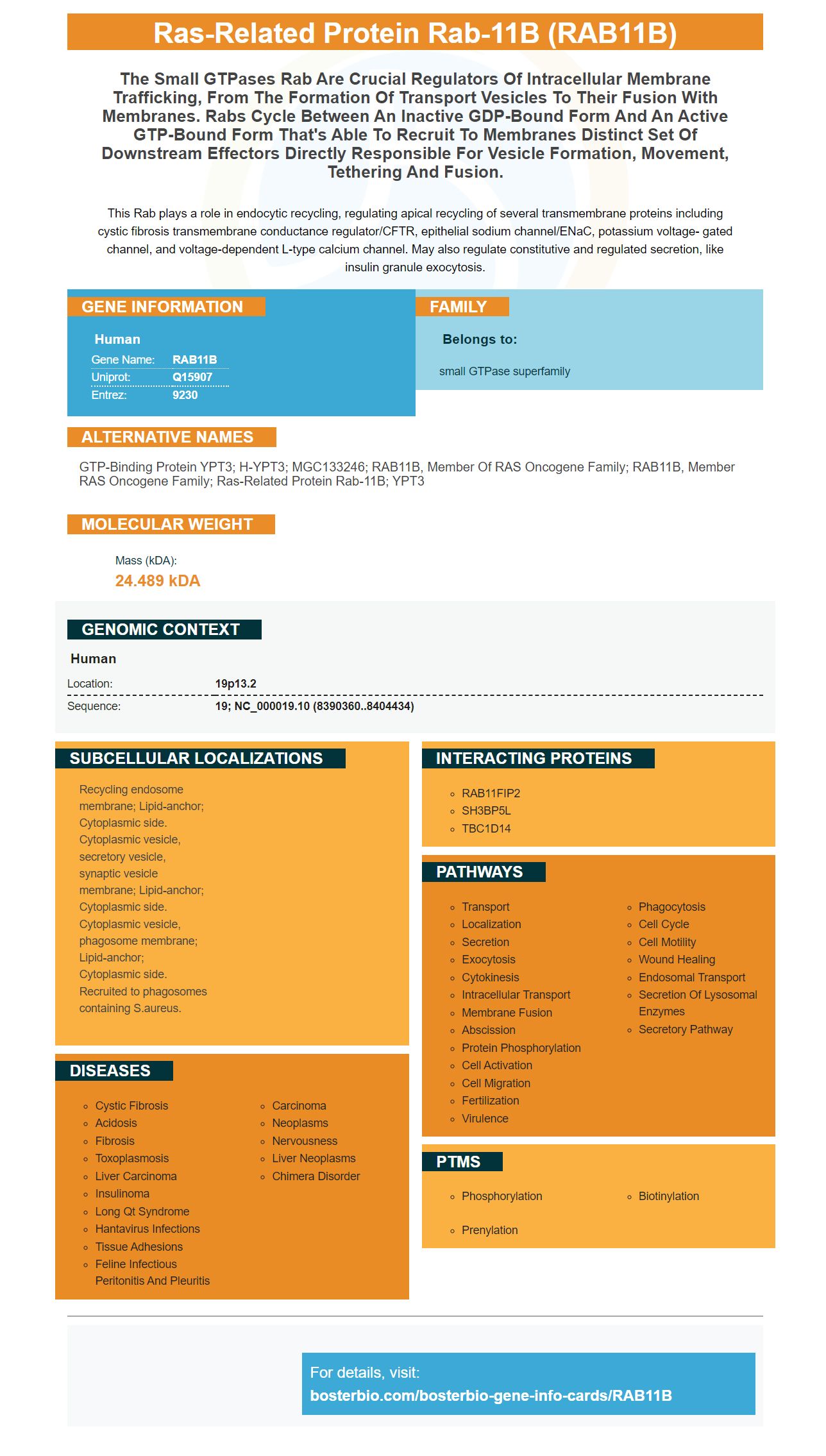
Facts about Ras-related protein Rab-11B.
This Rab plays a role in endocytic recycling, regulating apical recycling of several transmembrane proteins including cystic fibrosis transmembrane conductance regulator/CFTR, epithelial sodium channel/ENaC, potassium voltage- gated channel, and voltage-dependent L-type calcium channel. May also regulate constitutive and regulated secretion, like insulin granule exocytosis.
Human | |
---|---|
Gene Name: | RAB11B |
Uniprot: | Q15907 |
Entrez: | 9230 |
Belongs to: |
---|
small GTPase superfamily |
GTP-binding protein YPT3; H-YPT3; MGC133246; RAB11B, member of RAS oncogene family; RAB11B, member RAS oncogene family; ras-related protein Rab-11B; YPT3
Mass (kDA):
24.489 kDA
Human | |
---|---|
Location: | 19p13.2 |
Sequence: | 19; NC_000019.10 (8390360..8404434) |
Recycling endosome membrane; Lipid-anchor; Cytoplasmic side. Cytoplasmic vesicle, secretory vesicle, synaptic vesicle membrane; Lipid-anchor; Cytoplasmic side. Cytoplasmic vesicle, phagosome membrane; Lipid-anchor; Cytoplasmic side. Recruited to phagosomes containing S.aureus.
In this article, we'll discuss the usefulness of the Rab11a marker for identifying a bona fide internal exosome. We'll also explore its biological activities, including increasing Cav-1 secretion. In the end, we'll conclude that Rab11a is an invaluable marker for studying exosome biogenesis in Drosophila. Read on to discover more.
The discovery of a new type of human exosome, called Rab11a-exosomes, has opened a powerful new avenue in the study of these cellular vesicles. The study of the role of Rab11a in the formation of exosomes allows researchers to understand how genes control exosome formation. This type of exosome has been implicated in tumorigenesis, where the ability to control the tumor promoting growth, and possibly in immune system regulation.
The exact mechanism of exosome sorting is unknown, but the discovery of specific markers may facilitate better detection of cancer cells by recognizing these signatures in the body fluids. Many groups are developing routine blood tests to detect exosomes, and this research has already shown promising results. Rab11a and CD91 are known exosome antigens, and their expression levels vary according to the stage of cancer.
The new method of identifying exosomes will also allow for faster diagnosis of diseases. Because of the unique properties of exosomes, this approach is able to integrate several detection platforms, including acoustic and elastic lift force. Moreover, exosome isolation will be scalable and efficient. But it's still important to note that Rab11a is only one of the markers used to identify an exosome.
RAB11B, a receptor for the EGFR, is an important regulator of cellular function. It is required for a wide variety of biological functions, including protein synthesis, cell adhesion, apoptosis, apoptotic cell death, and the release of endosomes. Inhibiting these activities requires the ability to identify the exact location of Rab11.
Several studies have shown that Rab11 regulates membrane trafficking. Inhibiting Rab11 expression inhibits the return of the influenza hemagglutinin receptor to the cell surface. Moreover, Rab11 is involved in the trans-Golgi to plasma membrane transport. Rab11A and -B are the two major subunits that interact in the process of exosome secretion.
In Xenopus retina cell-free extracts, Rab11B-positive Rab cells contained immature rhodopsin. This is indicative of a defective transport of rhodopsin. The immature rhodopsin accumulated in Drosophila photoreceptors with dominant-negative Rab11N124I.
The Rab5/Rab11 system regulates many different cellular processes, including hemocyte proliferation and differentiation. Rab5 and Rab11 are involved in the control of multiple proteins involved in cellular signaling, including Eiger/JNK, Toll, and EGFR. By inhibiting Rab11 activity, researchers can investigate the cellular functions of these proteins. There are numerous potential applications for this marker.
There is a peptide that increases Cav-1 secretion from prostate cancer cells. This peptide is composed of amino acids 82 to 101, and is conjugated to the antennapedia protein transduction domain. This peptide is involved in regulating the function of eNOS, which inhibits the activity of Src. It also inhibits albumin-induced phosphorylation. These findings have implications for treatments of prostate cancer and BPH.
Phosphorylation of Cav1 affects vesicle trafficking. Specifically, phosphorylation of Cav1 results in increased spreading of the protein's negatively-charged N-terminal phosphotyrosine residues, which promote caveolae formation and release from the plasma membrane. In addition, it inhibits cav-2 expression. Although this peptide is not required for caveolae formation, it increases Cav-1 secretion.
Overexpression of Cav1 inhibits CT-b internalization in the absence of caveolae. It may also inhibit the internalization of dysferlin without direct interaction with the protein. Therefore, overexpression of Cav1 inhibits dysferlin internalization by indirect mechanisms, such as by modulating the protein content of cell surface rafts. The presence or absence of Cav1 modulates the dynamic equilibrium of cholesterol, lipids, and protein within rafts.
Y14F mutants significantly increased the efficiency of FRET when compared with Y14D mutant. The mutants formed fewer contacts than Y14D mutant. Y14F increased the efficiency of FRET by 48%. This mutant has reduced the number of mobile vesicles and the number of detachment events. It increases Cav-1 secretion in human vascular smooth muscle cells.
A recent study has found that the Y14 mutant rescues the uptake of caveolae-mediated albumin in Cav1-/ MLECs. The mutants expressed Myc-tagged Cav1-constructs to detect Myc-positive cells. In the uptake assays, Cav1-/-MLEC cells were stained with an anti-Myc antibody. The albumin-labeled BSA was washed with phosphate-free water before being used in the experiments.
The FRET efficiency of Cav1-tagged constructs was assessed. When Cav1 oligomerizes, it facilitates invagination of the plasma membrane. Moreover, Cav1 enhances the expression of VEGF and IL-6 genes in human pancreatic cancer cells. The experiments were repeated with YFP-CFP-tagged cells and compared with controls. These results indicate that Cav1-phosphorylation can promote the formation of caveolae.
SIMPLE, which stands for small integral membrane protein of the late endosome, was found to regulate the sorting of cargo into exosomes. It is a gene whose overexpression in the drosophila exosomes is a useful marker for exosome biogenesis in Drosophila. When SIMPLE is overexpressed in COS cells, it increases secretion of exosomes. However, this increased secretion occurred only when SIMPLE was mutated to disrupt proper MVB formation.
Recently, the exosome was thought of as a garbage bag. However, recent studies have shown that they have intriguing roles, and can be used as drug delivery systems and contain key molecules that play a role in tumour development. This review summarizes the latest research on exosome biomarkers. MicroRNAs are the more common molecules found inside exosomes. Improvements in techniques have made the study of exosomal RNA and protein a more feasible endeavor.
It has also been shown that small EVs do not carry the pHluorin tag. Furthermore, the pHluo-M153R-CD63 marker colocalized with classical markers of exosome biogenesis, including HSP70 and flotillin. Therefore, this protein is an excellent tool to study exosome biogenesis in Drosophila.
YKT6 has been shown to be a crucial factor in exosome biogenesis in two separate studies. Depletion of YKT6 decreased TSG101 in human lung cancer cells, while depletion of syntaxin 1A reduced the secretion of Evi-bearing exosomes. Syntaxin 1A, another Q-SNARE, has been shown to inhibit exosome biogenesis in Drosophila S2 cells.
Although exosomes are cell-specific, the release of these vesicles is regulated by different cellular conditions and by external factors. Different cells in Drosophila have different vesicles with different protein compositions. These differences in exosome release have been discovered using a method called ultracentrifugation. However, kinetic studies are not very common in the literature.
The study by Murrow et al. suggests that ATG12-ATG3 deficiency leads to impaired basal autophagy in Drosophila, but does not affect starvation-induced autophagy. Interestingly, exosome release is measured by a marker that measures the total amount of protein in exosomes. This suggests a link between exosomes and autophagy.
PMID: 7811277 by Zhu A.X., et al. Molecular cloning of two small GTP-binding proteins from human skeletal muscle.
PMID: 11495908 by Hales C.M., et al. Identification and characterization of a family of Rab11-interacting proteins.
*More publications can be found for each product on its corresponding product page