This website uses cookies to ensure you get the best experience on our website.
- Table of Contents
Facts about Ras-related protein Rab-10.
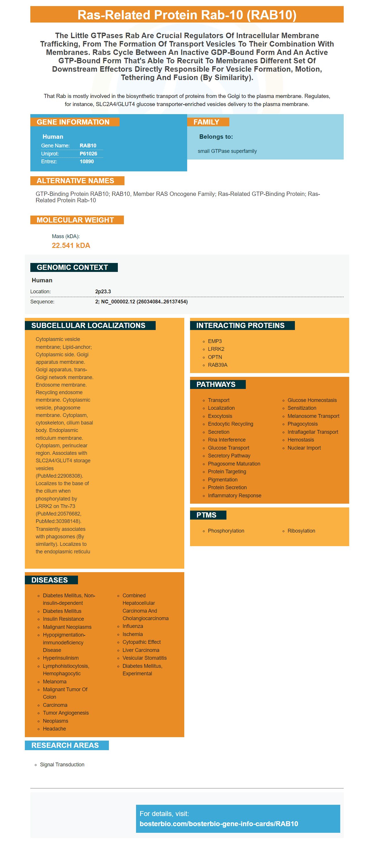
That Rab is mostly involved in the biosynthetic transport of proteins from the Golgi to the plasma membrane. Regulates, for instance, SLC2A4/GLUT4 glucose transporter-enriched vesicles delivery to the plasma membrane.
Human | |
---|---|
Gene Name: | RAB10 |
Uniprot: | P61026 |
Entrez: | 10890 |
Belongs to: |
---|
small GTPase superfamily |
GTP-binding protein RAB10; RAB10, member RAS oncogene family; ras-related GTP-binding protein; ras-related protein Rab-10
Mass (kDA):
22.541 kDA
Human | |
---|---|
Location: | 2p23.3 |
Sequence: | 2; NC_000002.12 (26034084..26137454) |
Cytoplasmic vesicle membrane; Lipid-anchor; Cytoplasmic side. Golgi apparatus membrane. Golgi apparatus, trans-Golgi network membrane. Endosome membrane. Recycling endosome membrane. Cytoplasmic vesicle, phagosome membrane. Cytoplasm, cytoskeleton, cilium basal body. Endoplasmic reticulum membrane. Cytoplasm, perinuclear region. Associates with SLC2A4/GLUT4 storage vesicles (PubMed:22908308). Localizes to the base of the cilium when phosphorylated by LRRK2 on Thr-73 (PubMed:20576682, PubMed:30398148). Transiently associates with phagosomes (By similarity). Localizes to the endoplasmic reticulu
Anti-RAB10 marker is a recombinant human RAB10 gene. It is useful in the detection of phosphorylated Rab10a & Rab8a proteins. It is available from Boster Bio. Continue reading to discover more about this protein. Here are some examples of its common uses. This protein is expressed by E. coli and comes with a His -Tag.
This antibody recognizes human RAB10 proteins recombinantly. Rab10 is an essential protein in the relocalization domain of MyoVa's globular tail. This antigen was designed to monitor the protein's activity. The rabbit polyclonal antibodies were produced against human RAB10.
The purified Rab10-Q63L was incubated at 27degC with Mst3 for 2 h and analyzed using NTAffinityAnalysis software. Phosphorylation of Rab10 was determined using the same protocol. Rab10 binding was detected using PhosTag gel electrophoresis. The Rab10 was then phosphorylated, and Mst3 was bound to it. Figure 2 shows the two proteins phosphorylation isotherms.
This antibody measures LRRK2-dependent phosphorylation of Rab10 in cells. The MSD assay measures total and phosphorylated Rab10 in cells. Positive control was the recombinant RAB10 proteins (pT73). The assay was also validated against mice lacking RRRK2.
LRRK2 was used as a peripheral biomarker, while pT73 was used. They were applied to PBMC lysates in a concentration-dependent manner to determine linearity. The ranges for LRRK2 and pT73 were 191.9 to 3070 ug/mL. While pT73 was more sensitive with a range between three to twelve ug/mL.
Researchers used this antibody to test pRab10 expression using HEK293T cells. Two shRNAs were used against Rab10, the pRab10Q63L, as well as pRab10. They used two shRNAs against Rab10, pRab10-Q63L and pRab10. A t test was performed in order to determine the significance.
pRab10 (full-length MyoVa) co-localize in pericentriolar membranes. The two proteins were found to be relocalized in pericentriolar, with the co-localization of pRab10 (recombinant MyoVa) and pRab10. Rab10 and its inhibitor RILPL2 did not co-locate in the same cell.
Anti-RAB10 marker is a recombinant human RAB10 antibodies. This antibody detects and blocks p-ARF-10 from the body's cells membranes. It inhibits endosome formation and is involved with regulating endocytic recycle. It regulates endosomal protein PI(4,5),P2 in the cells.
RILP is a phosphorylation-inducible kinase that binds LRRK2-phosphorylated Rab10 and MyoVa. RILPL2 helps in cargo enrichment during ciliogenesis. It can be found in the cilia. RILPL2-phosphorylated Rab10 redistributes Myosin V and the mother centriole after 30 minutes of severe starvation.
This antibody recognizes pRab10, MyoVa. MyoVa GTD can bind both p-Rab10 and MyoVa. Full-length MyoVa MyoVa MCherry bound Rab10; MyoVa DC bound Rab10 but less Rab10. These results indicate that the protective Haplotype regulates the stability protein in a specific cell type.
LRRK2, a kinase, is highly expressed on immune cells. LRRK2's expression in glial tissues is dependent on LRRK2, however endogenous LRRK2 phosphorylation of Rab10 was reduced in mouse microglia. pT73 levels of Rab10 were also higher in mouse microglia derived iPSC neurons.
Antibodies against human protein phosphorylated are also known as anti-phosphorylated Ra proteins. They can recognize the phosphorylated rat or mouse versions of this protein. These antibodies can also cross-react to the phosphorylated Rab10 or other phosphorylated substances.
Boster Bio RAB10 is a monoclonal anti-rabbit that recognizes phosphorylated Rab10 and phospho-Rab8a proteins in human PBMC. This antibody failed in its ability to detect phosphorylated Rab8a signals if there were no Rab proteins. Positive controls were HEK293T cell lines that overexpressed LRRK2 receptor R1441C.
The antibody is made up of a light chain CDR2 CDR2 sequence. This sequence is conserved in many species. This fragment is sequenced as xASxLAS. The light chain CDR2 sequence is derived from two or more Clone 5 or Clone 19 sequences. The CDR includes a nonconserved amino acid. Additionally, amino acids may be added at the position indicated with the x in the aligned order.
The antigens used in the assay are referred to as epitopes. These are the antigens that bind to the antibody with greater affinity or avidity than the nontargeting compounds. The non-target compound could be a nucleic, lipid, carbohydrate, or three-dimensional molecule.
The anti-phosphorylated RablO monoclonal anti-phosphorylated antibody contains an amino acids sequence that is 90% identical with the sequence of the molecule. The heavy chain can be variable. The antibody contains two or more amino acids in its heavy chain. The protein is immunogenic if it recognizes phosphorylated Rab8a and Rab10.
Phosphorylation Rab10 and Rab8a are reliable clinical markers for diagnosing autoimmune and inflammatory disorders. A biomarker for a specific protein, RablO, is phosphatrylated Rab10. It can be used to detect patients with LRRK2 pathogenic mutations (V122V-N1437H and R141C).
Boster Bio is the company you should turn to if your research requires ELISA kits or you wish to create your own. Boster Bio specializes only in high-sensitivity ELISAs and WB/IHC compatible antibodies. They also offer custom services. You can even have them delivered free to your doorstep! For more information, contact Boster Bio today. Here are some ways you can contact Boster Bio
Optimisation of dilution ratios and antibodies is a crucial part of immunohistochemistry. Boster Bio optimizes antibody concentrations to detect as many targets possible. Multiplex Immunohistochemistry services provide high throughput for screening and quick assay processing. With multiplex Immunohistochemistry, the company can detect multiple targets simultaneously. It is also compatible with a wide range tissues and cell types.
Boster supplies Enzyme-Linked Immunosorbent Assay kits (ELISA), to test for various antigens as well as proteins and cytokines. Boster's ELISA Kits are backed with 20 years of industry experience and the trust of over 10,000 scientists around world. Picokine ELISA kit are a great way to accelerate scientific discovery. They contain enough reagents for 96 tests and have been validated across many samples.
The company also offers multiplex immunohistochemistry services (IHC). These services can be purchased at a fair price. Boster Bio offers ELISA services, as well as multiplex immunohistochemistry (IHC). Boster Bio's multiplex IHC reagents are high-quality and provide high throughput screening of data as well as confirmation of results. Their products are reliable and efficient with over 5,000 articles in scientific journals.
PMID: 12450215 by He H., et al. Identification and characterization of nine novel human small GTPases showing variable expressions in liver cancer tissues.
PMID: 16641372 by Babbey C.M., et al. Rab10 regulates membrane transport through early endosomes of polarized Madin-Darby canine kidney cells.