This website uses cookies to ensure you get the best experience on our website.
- Table of Contents
2 Citations 7 Q&As


1 Citations 4 Q&As

1 Citations 16 Q&As


5 Citations 16 Q&As


Facts about Platelet-derived growth factor receptor beta.

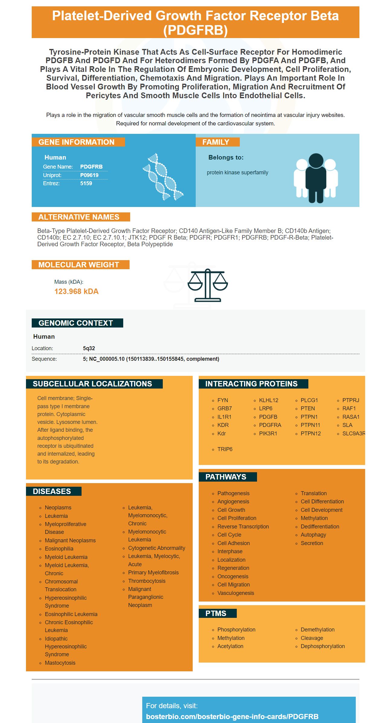
Plays a role in the migration of vascular smooth muscle cells and the formation of neointima at vascular injury websites. Required for normal development of the cardiovascular system.
| Human | |
|---|---|
| Gene Name: | PDGFRB |
| Uniprot: | P09619 |
| Entrez: | 5159 |

| Belongs to: |
|---|
| protein kinase superfamily |

beta-type platelet-derived growth factor receptor; CD140 antigen-like family member B; CD140b antigen; CD140b; EC 2.7.10; EC 2.7.10.1; JTK12; PDGF R beta; PDGFR; PDGFR1; PDGFRB; PDGF-R-beta; platelet-derived growth factor receptor, beta polypeptide
Mass (kDA):
123.968 kDA

| Human | |
|---|---|
| Location: | 5q32 |
| Sequence: | 5; NC_000005.10 (150113839..150155845, complement) |
Cell membrane; Single-pass type I membrane protein. Cytoplasmic vesicle. Lysosome lumen. After ligand binding, the autophosphorylated receptor is ubiquitinated and internalized, leading to its degradation.


Boster Bio’s PDGFRB Marker is what you want to know. This article discusses PDGFRB Marker, a transmembrane Glyprotein that promotes Angiogenesis. This marker is useful in many research applications. Continue reading for more information. This article also introduces CD146 which is a transmembrane protein that is also a biomarker and is often used in research.
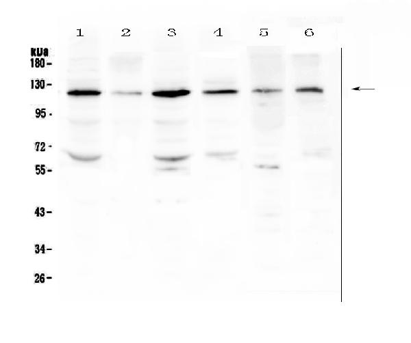
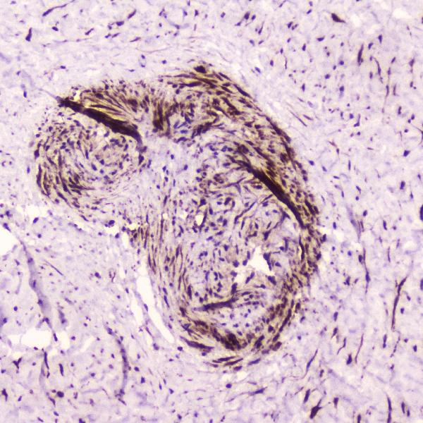
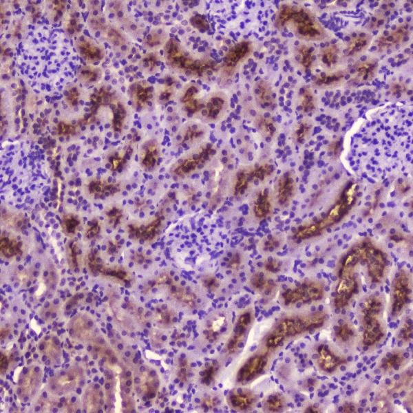
Boster Bio has developed the PDGFRB Marker, an antibody that can recognize human PDGF Receptor b. The antibody has been validated on both WB (and IHC) tests. It binds to the PDGF receptor B in human, mouse, and rat cells. It also reacts to other PDGF-receptors. Scientists can submit their results to Boster Bio for product credits.
It is not clear how PDGFRB contributes to angiogenesis. It is vital to the establishment of functional blood vessel and recruits mural cells into these developing structures. However, the exact function of PDGF/PDGFR-b in embryonic vascular development remains to be determined. For example, mice develop blood vessels around embryonic day 7.5, when the yolk sac is already filled with common hematopoietic/endothelial precursor cells.
A recent study found that a neutralizing anti-VEGFR-2 anti-PDGFRB antibody enhanced the antitumor effects in xenograft models of an antitumor anti-VEGFR-2 anti-VEGFR-2 antibody. Antibodies against PDGFRB can be used as an adjunct to anti-tumor or antiangiogenic agents. Its neutralizing antibody CP-673,451 inhibits PDGFRb expression in a variety of human tumors.
The study also showed that PDGFRB was expressed by both stromal as well ECs. However, PDGFRB expression was stronger in tumors than in perivascular cells. Furthermore, anti-PDGF-neutralizing antibodies could be useful to validate this hypothesis. Further research is needed in order to clarify the role that PDGFRB plays in angiogenesis.
We used E8 embryoid body to identify PDGFRB in embryonic renal cells. We fixed them in zinc buffer or in ethanol/acetone 1:1 (vol/vol) to identify PDGFRB. After blocking, we used antiPDGFR-b antibody for visualization of the coexpression of PDGFRb and CD31. Hoechst 33342 was also used to visualize the nuclei.
PDGF-B stimulates perivascular cells recruitment and is more prevalent in arterial and venous epithelium. Lack of PDGF-B inhibits the formation of small blood vessels and slows down cell proliferation. Mice that were deficient in PDGFB had decreased perivascular numbers and angiogenesis. In the laboratory, PDGFRb localized on the cords/tubes of angiogenic ECs and was elevated along with cord/tube formation. PDGFRb antibodies, which neutralize PDGFB, significantly reduced cord/tube production in aortic epithelial cell culture.
PDGFRa (and PDGFR-B) are expressed in breast cancer tumor stroma, connective tissue, and both sexes. Breast cancer cells invade bone to induce PDGFRb-b expression in the surrounding ECs. PDGFRB-deficient tumors have a greater MVD and angiogenic value than cancer cells expressing PDGFa.
Multiple myeloma can be treated by targeting the transmembrane regions of PDGFRB. The receptor binds to integrins and has two distinct functions. The receptor's TEL (ligand-binding) domain is the part that binds to integrins. The SAM domain, or pointed domain, mediates Oligomerization.
The PDGFR receptor activates when PDGF is present on the cell surface. This receptor is involved in a wide range cellular processes, including survival, proliferation, and cancer growth. It is expressed in fibroblasts and vascular smooth muscle cells. Various types of cells express the receptor. There are four types of the receptor: A, B and C. They bind to PDGF and stimulate their activation and activities.
The PDGFRbTM TM sequence contains one threonine and two serine residues. Threonine 545 is essential for productive interaction of the E5 protein. Both threonine and serine were lost in DTM14 and DTM21. TPb also includes GFP, a transmembrane protease.
PDGFRB, a transmemane glycoprotein, has a cell-surface tyrosine kinase region. It belongs to the platelet derived growth factor family of proteins that act as mitogens in cells of the mesenchymal origin. PDGFRb is flanked by G-M-C-stimulating factor receptor (GMCR) and the granulocyte-macrophage colony-stimulating factor receptor (G-M-CSF). The 5-q syndrome is believed to be caused by a translocation in the PDGFRB genes.
A recent study showed that a PDGFRB gene mutation called R561C compromises the autoinhibitory role of the JM domain. This mutation has been linked to a higher risk of myofibromas in tissues that have high PDGFRB signaling. Targeted signal transduction therapy (TST) may be used to help patients with this genetic mutation. This study highlights the role of PDGFRB in the pathogenesis of multiple myeloma and other disorders.
The PDGFRB genetic mutation has been associated with many cancers of blood-forming cell lines. Genetic rearrangements to the PDGFRB genome have also been linked to chronic eosinophilic lukemia. The most common translocation involves fusion of part of the PDGFRB gene with the ETV6 gene, which turns off its activity. Another gene can be fused with PDGFRB, which is called a "somatic mutation".
The PDGFRB gene is expressed in a wide variety of tissues and is implicated in numerous human diseases. Interestingly, however, the PDGFRB gene is poorly understood. This is due to a lack in published data regarding its biological function. Researchers have been able discover its potential for a variety of uses. We used two approaches to better understand this gene. First, RNA-Seq was used. The second method uses population RNA–Seq.
PMID: 2835772 by Gronwald R.G.K., et al. Cloning and expression of a cDNA coding for the human platelet- derived growth factor receptor: evidence for more than one receptor class.
PMID: 2850496 by Claesson-Welsh L., et al. cDNA cloning and expression of a human platelet-derived growth factor (PDGF) receptor specific for B-chain-containing PDGF molecules.
*More publications can be found for each product on its corresponding product page