This website uses cookies to ensure you get the best experience on our website.
- Table of Contents
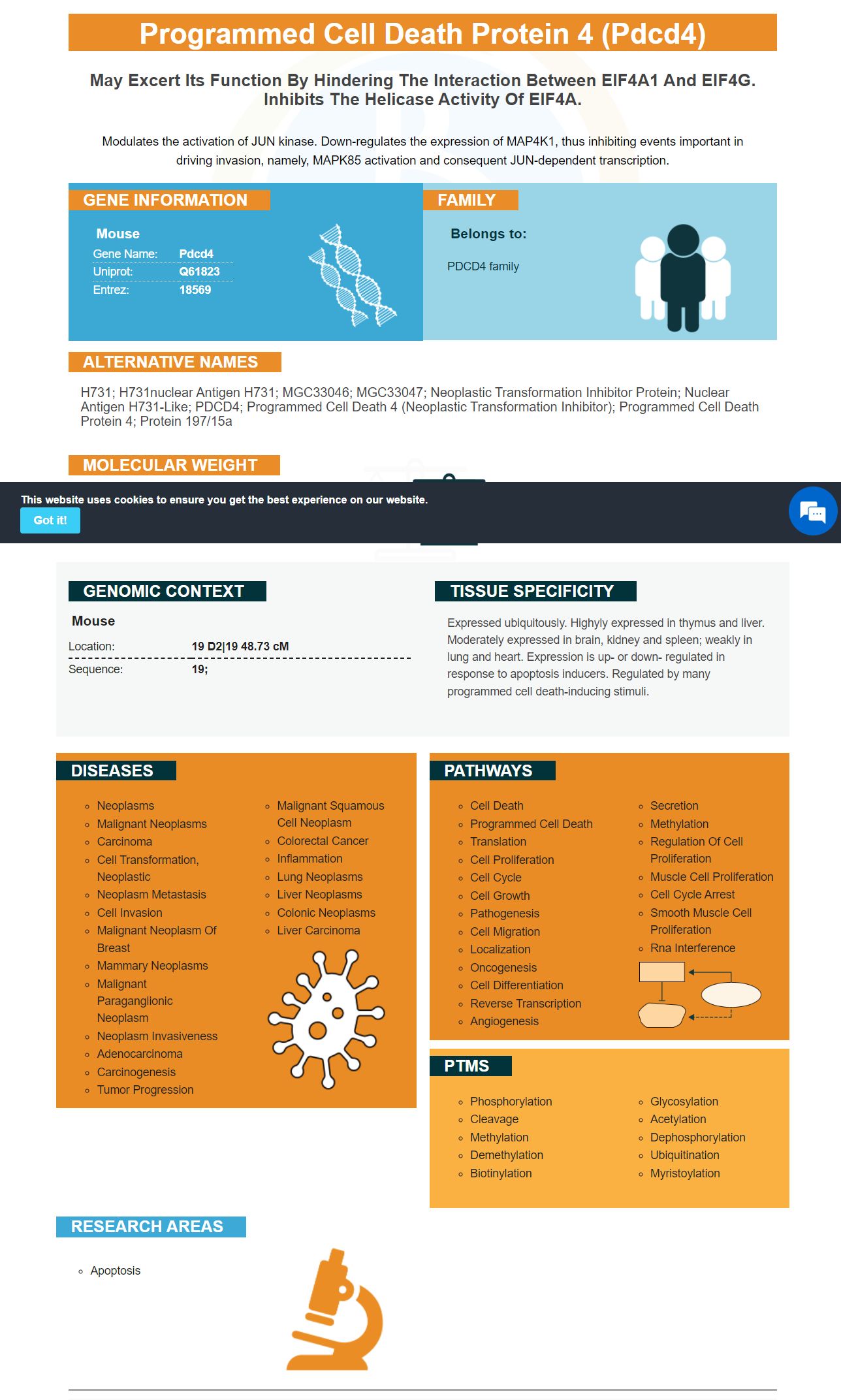
Facts about Programmed cell death protein 4.
Modulates the activation of JUN kinase. Down-regulates the expression of MAP4K1, thus inhibiting events important in driving invasion, namely, MAPK85 activation and consequent JUN-dependent transcription.
Mouse | |
---|---|
Gene Name: | Pdcd4 |
Uniprot: | Q61823 |
Entrez: | 18569 |
Belongs to: |
---|
PDCD4 family |
H731; H731nuclear antigen H731; MGC33046; MGC33047; Neoplastic transformation inhibitor protein; Nuclear antigen H731-like; PDCD4; programmed cell death 4 (neoplastic transformation inhibitor); programmed cell death protein 4; Protein 197/15a
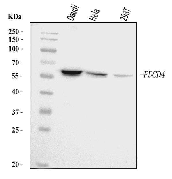
Mass (kDA):
51.702 kDA
Mouse | |
---|---|
Location: | 19 D2|19 48.73 cM |
Sequence: | 19; |
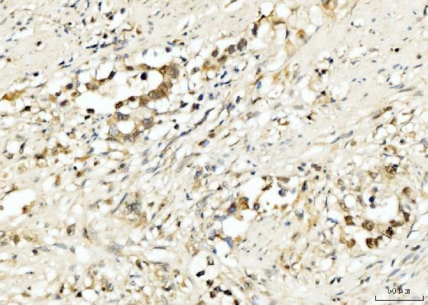
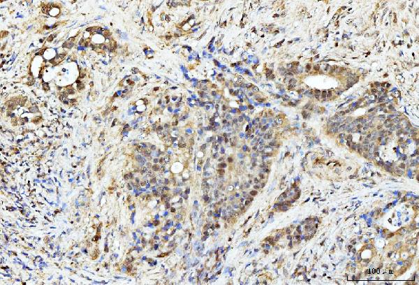
Expressed ubiquitously. Highyly expressed in thymus and liver. Moderately expressed in brain, kidney and spleen; weakly in lung and heart. Expression is up- or down- regulated in response to apoptosis inducers. Regulated by many programmed cell death-inducing stimuli.
The PDCD4 marker is an RNA polymerase chain reaction product that enables the measurement of mRNA expression levels of the PDCD4 protein. Using a real time PCR system equipped to use SYBR Green, PDCD4 levels were determined. To detect apoptosis, cells were stained with FITC-conjugated annexin V and propidium iodide.
The relative quantification technique was used to assess PDCD4 mRNA level in human lymphocytes. This method compares the levels of two target sequences in the same sample and expresses results as a ratio of the targets. The relative quantification method uses a reference gene (also called an endogenous control) that is expressed at constant copy numbers in all conditions. This method is especially useful in oncology and allows for the analysis of gene expression differences between samples.
A Roche LightCycler realtime-PCR system was used to extract total RNA from MCF-7 cells. This was then reverse-transcribed using the SuperScript III First Strand Synthesis Supermix Kit. A 20ml reaction containing cDNA in it was then done using SYBR Green I dye on a Light Cycler 480 II. Three independent experiments were carried out for each sample.
PDCD4 expression also inhibits lung cancer cell proliferation in H460 and A549 cells. Overexpression of this gene caused a decrease in HO-1 levels but had no effect upon NQQ and MRL levels. Also, the Nrf2 protein expression levels were identical to mRNA expression.
The cells expressing PDCD4 were co-transfected by FLAG-tagged Keap1 cells for 24 h. They were then cultured in the presence a reaction medium. The cells were tested for luciferase reporter activities using a colorimetric assay kit. Each experiment was done in duplicate.
The knockdown of PDCD4 reduces circ–NOL10 expression. In addition, down-regulation of miR-149-5p, miR-330-3p, and miR-450-5p decreased PDCD4 protein expression levels in breast cancer cells. This study has the potential for new targets to be identified in esophageal carcinogenesis.
PDCD4 over-expressing cells were transiently co-transfected with FLAG-tagged Keap1 and siRNA targeting Nrf2 protein. Luciferase activity in cells co-transfected with siRNA targeting p62 was determined by immunoblotting. Furthermore, flow cytometry was used for the measurement of caspase-3 activity.
Transfected cells were given a specific PCR primer in order to detect PDCD4 mRNA levels within human lymphocytes. These cells were cultured for 14 day to form visible colonies. The cells were then stained with 0.5% crystal violet. Three experiments were run to confirm the results. Cells were harvested following transfection to determine PDCD4 mRNA level.
The study found that silencing SNHG3 can inhibit cell proliferation and invasion in breast carcinoma cells. Similarly, PDCD4 mRNA expression levels were evaluated by SYBR Green incorporation on a Roche LightCycler real-time PCR system using a DDCT-Method. The results obtained from this experiment were compared with the reference gene by a MTT or EdU proliferation assay.
Cell apoptosis describes the process by which cells cease to exist. FITC-conjugated annexin V and propidium iodide staining were used to detect cell apoptosis in cultured human cancer cells. Both annexins recognizephospholipids, and have been proven to induce apoptosis. The ability of FITC-conjugated annexin 5 to bind to phospholipids was tested in two different natural sources. This was confirmed by western-blotting using antibiotin-HRP. The folding of annexinV was increased by the fusion between sfGFP, annexin IV and annexin 5.
For the annexin V and propidium-iodide staining, cells were harvested, centrifuged at low speed and resuspended in physiological washing buffer (PBS). The annexinV binding buffer contains calcium which is required for annexins to bind with PS. The cells were then added to a solution containing FITC-conjugated annexin V. The cells were stained with the dye and left for 20 minutes before being washed with PBS.
The detection of cell apoptosis using FITC conjugated antexinV and propidium iodate was a useful technique to identify the different types. FITC-conjugated annexin V binds phosphatidylserine (phosphatidylserine), which occurs in the inner leaflet of the cell membrane and is present in high concentrations during early apoptosis. Propidium Iodide, however, binds DNA but only enters cell membranes that are ruptured. This is characteristic of necrosis.
The FITC conjugated annaxinV staining and propidium ionide stains were used for cell apoptosi detection in cultured human tissues. HeLa cells were treated to an apoptotic stimulus at 0, 6, or 12 hours to check the specificity. The percentage of annexin V-positive cells is shown in the upper right-hand corner.
To determine whether apoptosis has been inducible in a specific type of cell, apoptosis must first be inducible. You can use UV radiation, 5 mM Act D or 200 ng/ml-1 antifas IgM antibodies. To ensure accurate results, a negative control should also been used. Next, remove 200 ml cells from each treatment. The cells should be stained with FITC conjugated annexin 5 and propidium iodide.
The FITC conjugated enzymatic process has been used to detect apoptosis of cells in vitro and vivo. Positive results have been obtained for imaging cell death with annexinV labeled using a radiotracer/PET probe. The same procedure can be used to detect membrane P in other organisms.
To determine if cells are undergoing apoptosis (or not), FITC-conjugated annexin V and PI were stained with a chromogenic color. The phospholipids in the microtiter plates were immobilized on polystyrene microtiter plate and protected by 5% skimmed Milk in HBS. The membrane protein was detected using an anti-biotin/-GFP antibody.
HO-1, an enzyme that is responsible for the conversion biliverdin/bilirubin, is known as HO-1. Its presence in the liver is thought be beneficial in many metabolic pathways and vascular reactive. HO-1 expression levels can also be related to inflammation, hyperoxia and even ischemia. This enzyme is believed play a critical role in antioxidant homeostasis. It is also responsible for preventing vascular injury.
The role of HO-1 in the development and progression of atherosclerosis remains largely unclear. Its antioxidant and anti-inflammatory properties may play an important role in the initial stages of atherosclerosis. Its antiapoptotic capabilities may be responsible for plaque rupture and lesion progression. HO-1 can also inhibit the growth of dendritic cells, regulate activity of phagocytes/macrophages, and regulate SMC proliferation.
Researchers compared tumors formed in mice after being injected with either wild type or HO-1-transfected melanomas. After four days, the tumors developed and were visible under the skin. The mice's survival time was also reduced by HO-1 overexpression. Despite the increased chance of developing melanoma in mice who had HO-1-overexpression, the mice lived a shorter time than their wild-type counterparts.
The study also revealed that HO-1 is an important mediator of angiogenesis. Both B16-WT and HO-1 melanoma cells expressed VEGF, a proangiogenic mediator. They released a comparable amount of VEGF in the medium. Hypoxic conditions also increased melanoma cell proliferation. This was in line with previous studies.
The researchers also examined the expression levels of HO-1 in a system-level approach to determine how oxPAPC affects this gene. Researchers also discovered genetic variations in the human population that altered the expression patterns these cells. They created a network of 11 gene co-expression modules using these data. HO-1 was associated with a number of pathways, including those involved in redox processes and sulfur amino acid metabolism.
To determine the expression of HO-1, researchers used a co-immunoprecipitation kit manufactured by Wuhan Boster Bio. In this method, total proteins were extracted using ice-cold lysis buffer containing 25 mM Tris-HCl (pH 8.0), 150 mM NaCl, and 1% SDS. Next, 500 ug was incubated for 4 hours at room temp with anti-SUMO-1. Finally, the immune complexes of antibodies were immobilized with pansorbin cells.
PMID: 8543179 by Shibahara K., et al. Isolation of a novel mouse gene MA-3 that is induced upon programmed cell death.
PMID: 8912629 by Onishi Y., et al. Molecular cloning of the genes suppressed in RVC lymphoma cells by topoisomerase inhibitors.